Physical Therapist's Guide to Patellofemoral Pain

What Is Patellofemoral Pain?

PFP may occur after a sudden increase in activities like running or jumping. Research suggests that PFP results from activity levels that are increased faster than the knee can adapt. Other contributing factors to PFP may include:

  • Weakness of the thigh muscles.

  • Specializing in a single sport, which requires repeating the same movements again and again.

  • Certain hip and knee coordination patterns during running and jumping activities.

PFP does not go away on its own. If you have symptoms of PFP, it’s important to seek care from a physical therapist so you can return to the activities that you enjoy.

How Does it Feel?

People with PFP may experience pain:

  • When walking up or down stairs or hills.

  • When playing a sport.

  • With deep knee bending (squatting).

  • When walking on uneven surfaces.

  • With activity, but improving with rest.

  • After sitting for long periods of time with the knee bent.

How Is It Diagnosed?

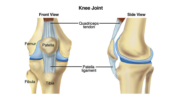
APTA-Knee-Joint-Patella-Pain_750x419.jpg

Your physical therapist will review your health history and conduct a series of tests to evaluate you and your knee. PFP is diagnosed by analyzing any movement that causes pain, and ruling out other possible conditions.

Your physical therapist may analyze your walking and running patterns. They may test the strength of your hip and thigh muscles to find out if weakness is contributing to your pain. Medical imaging, such as an X-ray or MRI, is not helpful in diagnosing PFP. However, your physical therapist may consult with an orthopedic physician who may order imaging to rule out other conditions.

How Can a Physical Therapist Help?

If PFP is diagnosed, your physical therapist will develop an exercise and rehabilitation program just for you. Your program may include:

Strengthening exercises. Your physical therapist will teach you exercises to help strengthen the muscles around the hip and the knee itself. Research shows that this type of exercise therapy is the best approach to managing PFP.

Taping. Your physical therapist may teach you how to apply tape to your knee, which may improve your ability to perform exercises that would normally be painful. However, taping alone will not resolve PFP. It must only be used along with your exercise program.

Shoe inserts. Your physical therapist may recommend shoe inserts to help reduce your pain when exercising. But inserts alone, like taping, will not treat PFP. Your physical therapist will design an exercise program to fit your specific needs and goals.

Coordination training. Based on your activity level, your physical therapist may help retrain your hip and knee movement patterns to reduce your knee pain.

This type of training is effective for athletes, in particular, and may focus on movements like:

  • Stair climbing.

  • Squatting.

  • Running and jumping.

Cross-training guidance. Physical therapists help athletes and active people perform different movements (cross-training). This helps them stay active until they can return to a favorite activity.

Return to full activity. Your physical therapist will help guide a gradual return to your favorite activities, such as running and jumping, and will teach you good overall exercise habits to help maximize the health of your knee.

Treatments That Do Not Work for PFP

While these can be appropriate for the treatment of other injuries or conditions, the following do notwork for PFP:

Quick fixes. “Passive” treatments like dry needling, ultrasound, laser, or electrical stimulation are not helpful for people with PFP. The most effective treatment for PFP is an exercise program that targets the hip and knee muscles.

Rest. If you are experiencing PFP, it is important to understand that rest only helps temporarily. Your pain will likely return when you go back to your normal activity. Rest is not helpful in the long term. A movement program guided by your physical therapist is your best treatment option.

Can this Injury or Condition be Prevented?

Current research shows that a person’s age, height, body weight, or foot alignment may not contribute to the risk of developing PFP at all. A knock-kneed posture also does not increase the risk of developing PFP.

However, a few preventive measures can be effective. To help reduce your risk of developing PFP:

  • Keep your thigh muscles strong.

  • Maintain good exercise habits.

  • Avoid rapid spikes in activity levels.

  • Participate in a variety of sports, rather than just repeating the same movements again and again.

What Kind of Physical Therapist Do I Need?

All physical therapists are prepared through education and experience to treat a variety of conditions or injuries including PFP. However, you may want to consider:

  • A physical therapist who is experienced in treating people with orthopedic, or musculoskeletal, problems.

  • A physical therapist who is a board-certified clinical specialist or who has completed a residency or fellowship in orthopedic physical therapy. This physical therapist will have advanced knowledge, experience, and skills that may apply to your condition.

  • While it may be tempting to seek quick fixes for your knee pain, there is no evidence that passive treatments work for persons with PFP. If you have PFP, seek care from a physical therapist who uses progressive exercise therapy for the treatment of this condition.

You can find physical therapists who have these and other credentials by using Find a PT, an online tool provided by the American Physical Therapy Association. You can search for physical therapists with specific clinical expertise in your geographic area.

General tips when you're looking for a physical therapist:

  • Get recommendations from family and friends or from other health care providers.

  • When you contact a physical therapy clinic for an appointment, ask about the physical therapist's experience in helping people with patellofemoral pain (PFP).

  • During your first visit with a physical therapist, you will be asked to describe your symptoms in as much detail as possible, and say what makes your symptoms worse. Here are some tips to prepare for your visit.

Further Reading

The American Physical Therapy Association (APTA) believes that consumers should have access to information that could help them make health care decisions and prepare for a visit with their health care provider.

The following articles provide some of the best scientific evidence related to physical therapy treatment of patellofemoral pain syndrome. The articles report recent research and give an overview of the standards of practice both in the United States and internationally. The article titles are linked either to a PubMed* abstract of the article or to free access of the full article, so you can read it or print out a copy to bring with you to your health care provider.

Patellofemoral pain: treating painful kneecaps. J Orthop Sports Phys Ther. 2019;49(9):633. Free Article.

Willy RW, Hoglund LT, Barton CJ, et al. Patellofemoral Pain. J Orthop Sports Phys Ther.2019;49(9):CPG1–CPG95. Free Article.

Neal BS, Lack SD, Lankhorst NE, Raye A, Morrissey D, van Middelkoop M. Risk factors for patellofemoral pain: a systematic review and meta-analysis. Br J Sports Med. 2019;53(5):270–281. Free Article.

Lack S, Barton C, Sohan O, Crossley K, Morrissey D. Proximal muscle rehabilitation is effective for patellofemoral pain: a systematic review with meta-analysis. Br J Sports Med. 2019;49(21):1365-1376. Free Article.

* PubMed is a free online resource developed by the National Center for Biotechnology Information (NCBI). PubMed contains millions of citations to biomedical literature, including citations in the National Library of Medicine’s MEDLINE database.

Authored by Christopher Bise, PT, DPT, MS. Revised and reviewed by Richard Willy, PT, PhD.

Meniscus Tear

Meniscal tears are common injuries to the cartilage of the knee that can affect athletes and nonathletes alike. These tears can be either “acute,” meaning they happen as a result of a particular movement, or “degenerative,” meaning they happen over time. Your physical therapist can help you heal a meniscal tear and restore your strength and movement. If surgery is required, your physical therapist can help you prepare for the procedure and recover following surgery.

Unknown.jpeg


What is a Meniscal Tear?

The meniscus is a cartilage disc that cushions your knee. Each of your knees has 2 menisci (plural of meniscus); one on the inner (medial) part of the knee, and the other on the outer (lateral) part of the knee. Together, they act to absorb shock and stabilize the knee joint.

Meniscal tears can be classified in 2 ways: acute or degenerative. An acute meniscal tear typically is caused by twisting or turning quickly on a bent knee, often with the foot planted on the ground. This mechanism of injury often produces related injuries, such as an ACL tear. Degenerative meniscal tears occur over time, due to repetitive stress being put on the knee, such as in a job or sport that requires a lot of squatting.


How Does it Feel?

When you tear the meniscus, you might experience:

  • A sharp, intense pain in the knee area

  • A "pop" or a tearing sensation in the knee area (acute injury)

  • Swelling within the first 24 hours of injury

  • Difficulty walking or going up or down stairs because of pain or a "catching or locking" sensation in the knee

  • Difficulty straightening or bending the knee fully


How Is It Diagnosed?

Your physical therapist will:

  • Conduct a thorough evaluation that includes a detailed review of your injury, symptoms, and health history.

  • Perform special tests to measure the range of motion (amount of movement) in your knee and determine which specific movements and positions increase your symptoms.

  • Use a series of tests that apply pressure to the meniscus to determine whether it appears to be damaged.

The results of these tests may indicate the need for further diagnostic tests—such as magnetic resonance imaging (MRI)—or a referral to an orthopedic surgeon for consultation.


How Can a Physical Therapist Help?

Meniscal tears can often be managed without surgery. A short course of treatment provided by a physical therapist can help determine whether your knee will recover without surgery. Your physical therapist can help control pain and swelling in the knee area and work with you to restore full strength and mobility to your knee. Your treatment may include:

Manual therapy. Your physical therapist may apply manual therapy—hands-on treatment that may include massage, stretching, or joint mobilization—to help reduce swelling and stiffness, and restore muscle function around the knee.

Icing. Your physical therapist will apply ice packs to the knee to help control any pain and swelling, and may instruct you to apply icing at home. Swelling is an important "guide" during your rehabilitation and can indicate your level of ability and recovery. If you experience an increase in swelling, your physical therapist will modify your treatment program or activity level to ensure your safest, most effective recovery.

Compression. Your physical therapist may recommend the use of compression bandages, stockings, or pumps to assist in the reduction of or prevent further accumulation of edema (swelling). Your physical therapist may include them as part of your regular treatments and teach you and your family how to use them at home.

NMES. Your physical therapist may use a treatment called neuromuscular electrical stimulation (NMES). NMES uses electrical current to gently stimulate/contract the muscles around your knee to help improve their strength.

Assistive devices. It may be necessary to use assistive devices such as crutches, a cane, or a walker in the short term. Your physical therapist will make recommendations about which device is best for you and will instruct you in how to use it properly.

Strengthening exercises. Your physical therapist will design exercises to build and maintain your strength during recovery and help restore full movement to the knee. You will be given a home program of exercises that are specific to your condition. Strengthening the muscles around the knee and throughout the leg helps ease pressure on the healing knee tissue.

Fitness counseling. As you recover, your physical therapist will advise you on ways to improve and maintain your fitness and activity levels, and will help you decide when you are ready to return to full activity.

If Surgery Is Required

Patients with more serious meniscal tears, or those who don’t respond to a course of physical therapy, may need surgery to repair the injured knee. Surgically removing the torn cartilage (a procedure called a meniscectomy) usually is a simple procedure that requires a course of physical therapy treatment following surgery. Many people are able to return to their previous level of activity, including sports, after approximately 4 months of treatment.

Meniscus removal. Following a simple meniscectomy, your rehabilitation will likely be similar to that for nonsurgical meniscal injuries. Your physical therapist may use ice and compression to control pain and swelling and will show you how to use these treatments at home. The focus of your treatment will be on helping you get back your strength and movement through a series of exercises performed in the clinic and at home, as well as with hands-on treatment (manual therapy). Initially, it is likely that you will need to use crutches or a cane for walking. Your physical therapist will help guide you in gradually placing your weight on the knee to stand or walk, to allow the meniscus and other tissue in the knee joint to slowly adjust to increased pressure.

Meniscus repair. Sometimes your surgeon will decide that the torn meniscus can be repaired instead of removed. Research studies show that if a meniscal repair is possible, it can reduce the risk of arthritis developing later in life. Rehabilitation following a meniscal repair is slower and more extensive than with meniscal removal because the repaired tissue must be protected while it is healing. The type of surgical technique performed, the extent of your injury, and the preferences of the surgeon often determine how quickly you will be able to put weight on your leg, stop using crutches, and return to your previous activities.

Following surgery for meniscal repair, your physical therapist will:

  • Help you control pain and swelling

  • Guide you through progressive reloading of weight to the knee to allow the cartilage to slowly adjust to increased compressive stress and pressure

  • Help restore your knee and leg range of motion

  • Teach you exercises to help restore your muscle strength

Return to Activity

Whether your torn meniscus recovered on its own or required surgery, your physical therapist will play an important role in helping you return to your previous activities. Your physical therapist will help you learn to walk without favoring the leg and perform activities like going up and down stairs with ease.

Return to work. If you have a physically demanding job or lifestyle, your physical therapist can help you return to these activities and improve how you perform them.

Return to sport. If you are an athlete, you may need a more extensive course of rehabilitation. Your physical therapist will help you fully restore your strength, endurance, flexibility, and coordination to help maximize your return to sports and prevent reinjury. Return to sport varies greatly from one person to the next and depends on the extent of the injury, the specific surgical procedure, the preference of the surgeon, and the type of sport. Your physical therapist will consider these factors when designing and adjusting your rehabilitation program, and will work closely with your surgeon to help decide when it is safe for you to return to sports and other activities.

.

Can this Injury or Condition be Prevented?

There is little research at this time to support exercise or other interventions such as bracing for prevention of meniscal injuries. However, you can make choices to help improve your overall fitness and keep your knees as strong and as healthy as possible. Practices that can help keep your knees strong include:

  • Regular exercise to help strengthen the muscles that support your knees

  • Staying physically active to prepare your body for the demands of a sport or strenuous activity

  • Avoiding twisting or turning quickly while your foot is planted on the ground, to help prevent stress to the knee that can cause a meniscal tear

If you already have knee problems, your physical therapist can help you develop a fitness program that takes your knees into account. Some exercises are better than others for those with a history of knee pain. Many exercises can be modified to fit your specific needs.


Real Life Experiences

Beau is a college sophomore who plays on his intramural lacrosse team. During a recent practice game, Beau twisted his left knee while performing a sharp turn to make a catch. He immediately heard a “pop” and felt pain in his knee joint. He was helped off the field by teammates and led back to his room, where he applied ice and rested for the rest of the evening.

The next morning, Beau felt pain when he put weight on his leg to get out of bed, and found he had difficulty walking; he also noticed some swelling on the inside of his knee. His roommate is in the physical therapy program at his university; he suggested Beau see a physical therapist.

Beau’s physical therapist gets his medical history and asks him to describe what happened in the game to get a sense of what might have happened to his knee. She then performs some tests that include movements that selectively stress the tissues of the knee to see if the symptoms can be provoked. She tells Beau that his symptoms may indicate a meniscal tear. She recommends that he consult with an orthopedic surgeon, who orders magnetic resonance imaging (MRI). The surgeon confirms a diagnosis of a medial meniscal tear. After consultation with the surgeon, Beau chooses to have the tear “cleaned up,” and have a small piece of the meniscus removed—a procedure called a meniscectomy.

Prior to surgery, Beau works with his physical therapist, who prescribes exercises and manual therapy to reduce the swelling, improve the knee range of motion, and restore muscle function around the knee—treatments that have been shown to improve surgical outcomes.

Following his surgery, Beau’s physical therapist controls the swelling around the knee joint with ice, and shows Beau how to ice his knee at home. She applies electrical stimulation to speed the recovery of the quadriceps muscle. She teaches him range-of-motion exercises and tells him how often he should be doing them at home. She teaches him how to use crutches to avoid putting pressure and weight on the knee while its tissues heal.

After 1 week, Beau is able to walk without his crutches, but has difficulty bending his knee fully or straightening it when he walks. His physical therapist works with him on improving his walking pattern, and continues to address his knee range of motion, strength, swelling, and pain. His exercises are adjusted as he heals to continue to challenge him and move his recovery forward.

After 3 weeks, Beau has full range of motion and increased strength in his involved leg. He has good balance and no pain while walking. His physical therapist guides him as he returns to jogging and then running. She gives Beau feedback on how much he should be running, how running should feel, and what to do to ensure a symmetrical running pattern to keep his knee and his other joints safe.

At 4 weeks, Beau’s treatment progresses to sports-related rehabilitation activities, which include moving quickly from side to side and learning how to safely turn to field a catch. His physical therapist provides guidance and training to help Beau avoid reinjury. Beau reports his leg muscles have gained strength, and he feels more stable as he executes his movements.

After 6 weeks of treatment, Beau rejoins his team for a playoff game and, with newfound confidence, sets up his teammate for a winning goal!

This story was based on a real-life case. Your case may be different. Your physical therapist will tailor a treatment program to your specific case.


What Kind of Physical Therapist Do I Need?

All physical therapists are prepared through education and experience to treat a variety of conditions or injuries. You may want to consider:

  • A physical therapist who is experienced in treating people with orthopedic (musculoskeletal) problems or sports injuries.

  • A physical therapist who is a board-certified orthopaedic clinical specialist or who completed a residency or fellowship in orthopedic physical therapy or sports physical therapy has advanced knowledge, experience, and skills that may apply to your condition.

You can find physical therapists who have these and other credentials by using Find a PT, the online tool built by the American Physical Therapy Association to help you search for physical therapists with specific clinical expertise in your geographic area.

General tips when you're looking for a physical therapist:

  • Get recommendations from family and friends or from other health care providers.

  • When you contact a physical therapy clinic for an appointment, ask about the physical therapist's experience in helping people with a meniscal tear.

During your first visit with the physical therapist, be prepared to describe your symptoms in as much detail as possible, and say what makes your symptoms worse.


Further Reading

The American Physical Therapy Association (APTA) believes that consumers should have access to information that could help them make health care decisions and also prepare them for their visit with their health care provider.

The following articles provide some of the best scientific evidence related to physical therapy treatment of meniscal injuries. The articles report recent research and give an overview of the standards of practice for treatment both in the United States and internationally. The article titles are listed by year and are linked either to a PubMed* abstract of the article or to free access of the full article, so that you can read it or print out a copy to bring with you to your health care provider.

Beaufils P, Pujol N. Management of traumatic meniscal tear and degenerative meniscal lesions: save the meniscus. Orthop Tramatol Surg Res. 2017 September 2 [Epub ahead of print]. doi: 10.1016/j.otsr.2017.08.003. Article Summary in PubMed.

Moses MJ, Wang DE, Weinberg M, Strauss EJ. Clinical outcomes following surgically repaired bucket-handle meniscus tears. Phys Sportsmed. 2017 May 23 [Epub ahead of print]. doi: 10.1080/00913847.2017.1331688. Article Summary in PubMed.

Skou ST, Lind M, Holmich P, et al. Study protocol for a randomised controlled trial of meniscal surgery compared with exercise and patient education for treatment of meniscal tears in young adults. BMJ Open. 2017;7(8):e017436. Free Article.

Hare KB, Stefan Lohmander L, Kise NJ, et al. Middle-aged patients with an MRI-verified medial meniscal tear report symptoms commonly associated with knee osteoarthritis. Acta Orthop. 2017 August 8 [Epub ahead of print]. doi: 10.1080/17453674.2017.1360985. Free Article.

Katz JN, Brophy RH, Chaisson CE, et al. Surgery versus physical therapy for a meniscal tear and osteoarthritis. N Engl J Med. 2013;368:1675-1684. Article Summary in PubMed.

Logerstedt DS, Snyder-Mackler L, Ritter RC, Axe MJ. Knee pain and mobility impairments: meniscal and articular cartilage lesions. J Orthop Sports Phys Ther. 2010;40(6):A1-A35. Free Article.

Heckmann TP, Barber-Westin SD, Noyes FR. Meniscal repair and transplantation: indications, techniques, rehabilitation, and clinical outcome. J Orthop Sports Phys Ther. 2006;36:795-814. Article Summary in PubMed.

Lowery DJ, Farley TD, Wing DW, et al. A clinical composite score accurately detects meniscal pathology. Arthroscopy.2006;22:1174-1179.  Article Summary in PubMed.

Fitzgerald GK, Piva SR, Irrgang JJ. A modified neuromuscular electrical stimulation protocol for quadriceps strength training following anterior cruciate ligament reconstruction. J Orthop Sports Phys Ther. 2003;33:492-501. Article Summary in PubMed.

*PubMed is a free online resource developed by the National Center for Biotechnology Information (NCBI).  PubMed contains millions of citations to biomedical literature, including citations in the National Library of Medicine’s MEDLINE database.

 Authored by Christopher Bise, PT, DPT, MS. Reviewed by the MoveForwardPT.com editorial board.


Hip Impingement (Femoroacetabular Impingement)

Hip impingement involves a change in the shape of the surface of the hip joint that predisposes it to damage, resulting in stiffness and pain. Hip impingement is a process that may precede hip osteoarthritis. It most often occurs in young, active people. A recent study found that 87% of teens and adults with hip pain showed evidence of hip impingement on diagnostic images taken of their hip joints. To treat hip impingement, physical therapists prescribe stretches and strengthening exercises to better balance the muscles around the hip to protect it, and use manual therapies to help restore range of motion and increase comfort.

What is Hip Impingement?

There are 2 types of hip impingement; they may occur alone or together.

Pincer-Type Impingement

  • In pincer-type impingement, the hip socket (acetabulum), which is usually angled forward, may be angled toward the back, or protruding bone may be present on the pelvis side of the hip joint making the socket a deeper recess that covers more of the ball or head of the femur bone.

  • The overgrown bone or incorrect angle of the socket causes the labrum, a rim of connective tissue around the edge of the hip socket, to be pinched. Over time, this extra pressure to the labrum when flexing (moving the leg forward) leads to wear and tear that can cause inflammation and could result in a tear. If this condition persists, eventually the cartilage that lines the hip joint can become worn and form holes.

  • This condition affects men and women equally; symptoms often begin early, appearing at any time between 15 to 50 years of age.


Cam-Type Impingement

  • In cam-type impingement, the shape of the bone around the head of the femur—the ball at the top of the bone in the thigh—is misshapen. It can vary from the normal round ball shape, or have overgrown bone formed at the top and front. The nickname “pistol grip” deformity is given to the appearance of the bony overgrowth on x-rays.

  • The overgrown or misshapen bone contacts the cartilage that lines the hip socket, and can cause it to peel away from the bone in the socket. The labrum can become worn, frayed, or torn as well.

  • This condition affects men to women at a ratio of 3 to 1; symptoms often manifest during the teen years and 20s.

HipImpingement-SM.jpg

Signs and Symptoms

Hip impingement may cause you to experience:

  • Stiffness or deep aching pain in the front or side of the hip or front of the upper thigh while resting.

  • Sharp, stabbing pain when standing up from a chair, squatting, rising from a squat, running, "cutting," jumping, twisting, pivoting, or making lateral motions.

  • Hip pain described in a specific location by making a "C" with the thumb and hand and placing it on the fold at the front and side of the hip, known as the "C-sign."

  • Pain that most often starts gradually, but can also remain after another injury resolves.

  • Pain that increases with prolonged sitting or forward leaning.

  • Feeling less flexible at the hips, including a decreased ability to turn your thigh inward on the painful side.

How Is It Diagnosed?

Your physical therapist will evaluate the range of motion (movement) of the hip and surrounding joints, and test the strength of the muscles in that area. Your therapist will feel the hip joint and surrounding muscles to evaluate their condition. The examination will include observing how you move, standing from a sitting position, walking, running, or squatting, as appropriate. Your physical therapist may perform special tests to help determine whether the hip is the source of your symptoms. For instance, the therapist may gently roll your leg in and out (the “log roll” test), or bend your hip up and in while turning the lower leg out to the side (the "FADDIR" test) to assess your condition.

If further diagnosis is needed, your doctor may order diagnostic tests to help identify any joint changes, including x-rays, magnetic resonance imaging (MRI), or diagnostic injections. Hip impingement can occur at the same time as low back, buttock, or pelvic pain, or from conditions such as bursitis or groin strain. The final diagnosis of hip impingement may take some time, especially when other conditions are present.

How Can a Physical Therapist Help?

Without Surgery

When an active person develops hip pain, but does not have severe symptoms or joint damage, the recommended treatment is physical therapy. The following interventions can help decrease pain, improve movement, and avoid the progression of hip impingement and the need for surgery:

  • Improving the strength of your hips and trunk. Strengthening of the hips and trunk can reduce abnormal forces on the already injured joint and help with strategies to compensate.

  • Improving hip muscle flexibility and joint mobility. Stretching tight muscles can reduce abnormal forces that cause pain with motion. Joint mobilization may help ease pain from the hip joint; however, these treatments do not always help range of motion, especially if the shape of the bone at the hip joint has changed.

  • Improving tolerance of daily activities. Your physical therapist can consider your job and recreational activities and offer advice regarding maintaining postures that are healthier for your hip and activity modification. Often this involves limiting the amount of bending at the hip to avoid further hip damage.


Following Surgery

Surgery for hip impingement is performed with arthroscopy. This is a minimally invasive type of surgery, where the surgeon makes small incisions in the skin and inserts pencil-sized instruments into the joint to repair damage. The surgeon may perform 1 or several techniques during your procedure as needed. The surgeon may remove or reshape the bone on the pelvis or femur side of the joint, and repair or remove the damaged labrum or cartilage of the hip joint.

Postsurgical physical therapy varies based on the procedure performed. It may include:

  • Ensuring your safety as you heal. Your physical therapist may recommend that you limit the amount of weight you put on the operated leg if there was a repair of the labrum. You may wear a brace to help limit the amount of bending at the hip. You might also use crutches to avoid overloading the leg if the bone on the femur was reshaped.

  • Improving your range of motion, strength, and balance. Your physical therapist will guide you through safe range-of-motion, strengthening, and balance activities to improve your movement as quickly as possible while allowing the surgical site to heal properly.

  • Instructions on returning to an active lifestyle. Most people return to normal daily activities about 3 months after surgery, and to high-level activities and sports 4 to 6 months after surgery. Your physical therapist will recommend a gradual return to activity based on your condition—research shows that 60% to 90% of athletes return to their previous playing ability depending on the surgical procedure performed and the sport.

Can this Injury or Condition be Prevented?

Currently there are no recommendations to prevent hip impingement. Despite a major increase in research to learn more about hip impingement, there is a great deal that is unknown. For instance, many active young people whose x-rays show hips as being abnormal do not have pain despite continuing to live active lives and participate in sports.

However, there is evidence that physical therapy interventions along with anti-inflammatory drugs can decrease pain, slow joint damage, and improve function. This is particularly important in those with mild hip impingement, those who are attempting to avoid surgery, and those who are not candidates for surgery.

Real Life Experiences

Lindsay is an active high school senior who plays shortstop for her school's softball team. Over the last several months, she has had progressively worsening pain on the front and side of her left hip. It started as an occasional sharp pain when she fielded ground balls at practice, and it eventually developed into aching and stiffness of the hip while resting. Lindsay occasionally develops hip pain while sitting in class or at the movies. In the past couple of weeks, she has found it hard to lean forward to tie her shoes. Her mom has been worrying about her pain and takes Lindsay to her physical therapist.

At the evaluation, the physical therapist finds that Lindsay has weakness around her hip and trunk muscles, decreased hip mobility, pain when flexing the hip, pain returning to a standing position after squatting, and decreased balance when standing on her affected leg. Her physical therapist diagnoses mild hip impingement in her left hip. Lindsay sees her physical therapist 1-2 times a week for the next 6 weeks.

Her treatments focus on developing a home program for strengthening her hips and trunk, and the therapist uses manual therapy for the hip to improve her comfort and allow her to perform more activities. The therapist works with Lindsay to change how she moves when standing from a seated position, and also to modify how she moves when playing the infield in softball. Lindsay also spends less time in the positions that bother her hip in the weight room and on the practice field, following recommendations from her physical therapist. After 3 weeks, the majority of her pain has subsided, and by 6 weeks, she is playing in games pain-free.

Lindsay meets her goal of finishing her senior year with the softball team. However, she is considering other ways to stay active after she graduates that don’t involve bending forward as much.

This story was based on a real-life case. Your case may be different. Your physical therapist will tailor a treatment program to your specific case.

What Kind of Physical Therapist Do I Need?

All physical therapists are prepared through education and experience to treat patients who have hip impingement. You may want to consider:

  • A physical therapist who is experienced in treating people with musculoskeletal problems. Some physical therapists have a practice with a sports or orthopaedic focus.

  • A physical therapist who is a board-certified clinical specialist or who completed a residency or fellowship in sports or orthopaedic physical therapy. This therapist has advanced knowledge, experience, and skills that may apply to your condition.

You can find physical therapists who have these and other credentials by using Find a PT, the online tool built by the American Physical Therapy Association to help you search for physical therapists with specific clinical expertise in your geographic area.

General tips when you're looking for a physical therapist (or any other health care provider):

  • Get recommendations from family and friends or from other health care providers.

  • When you contact a physical therapy clinic for an appointment, ask about the physical therapists' experience in helping people with hip impingement.

  • During your first visit with the physical therapist, be prepared to describe your symptoms in as much detail as possible, and say what makes your symptoms worse.

Further Reading

The American Physical Therapy Association (APTA) believes that consumers should have access to information that could help them make health care decisions and also prepare them for their visit with their health care provider.

The following articles provide some of the best scientific evidence related to physical therapy treatment of problems related to hip impingement. The articles report recent research and give an overview of the standards of practice for treatment both in the United States and internationally. The article titles are linked either to a PubMed* abstract of the article or to free full text, so that you can read it or print out a copy to bring with you to your health care provider.

Byrd JW. Femoroacetabular impingement in athletes, part I: cause and assessment. Sports Health. 2010;2:321-333. Free Article.

Byrd JW. Femoroacetabular impingement in athletes, part II: treatment and outcomes. Sports Health. 2010;2:403-409. Free Article.

Enseki KR, Martin RL, Draovitch P, et al. The hip joint: arthroscopic procedures and postoperative rehabilitation. J Orthop Sports Phys Ther. 2006:36:516-525. Article Summary in PubMed.

*PubMed is a free online resource developed by the National Center for Biotechnology Information (NCBI). PubMed contains millions of citations to biomedical literature, including citations in the National Library of Medicine’s MEDLINE database.

Authored by Jennifer Miller, PT, MPT, SCS. Reviewed by the MoveForwardPT.com editorial board.
Greater Trochanteric Bursitis

Greater trochanteric bursitis (GTB) is an irritation of the bursa, a fluid-filled sac that sits on top of the greater trochanter, a bony prominence on the outside of the hip bone (femur). The bursa acts as a cushion to decrease friction between the outside of the hip bone and muscles attaching to the bone; bursitis results when the bursa on the outside of the hip bone becomes irritated. Greater trochanteric pain syndrome is the term used when the condition also includes irritation to the tendons of the gluteal muscles that sit beneath the bursa. 

Most often, GTB is the result of repetitive friction to the bursa due to a combination of muscle weakness and tightness affecting the outside of the hip. The condition is most often treated with physical therapy to restore normal function.

GTB may result from a combination of several different variables, including:

  • Gluteal muscle weakness

  • Iliotibial (IT) band (a thick band of tissue that runs along the outside of the leg from the pelvis to the knee) tightness

  • Hip muscle tightness

  • Abnormal hip or knee structure

  • Abnormal hip or knee mechanics (movement)

  • Improper movement technique with repetitive activities

  • Change in an exercise routine or sport activity

  • Improper footwear


images-1.jpeg


How Does it Feel?

People with GTB may experience:

  • Tenderness to touch on the outside of the hip

  • Pain that can vary from sharp to dull, and can radiate to the buttock, groin, thigh, or knee

  • Pain that is intermittent and symptomatic for a prolonged period

  • Pain when lying on the involved side

  • Pain and stiffness with prolonged sitting, walking (worst with the first few steps), negotiating stairs, or squatting

  • Pain that may increase during prolonged activity


How Is It Diagnosed?

The goals of the initial examination are to assess the degree of the injury, and determine the cause and contributing factors to it. GTB is a condition that develops as a consequence of repetitive irritation in the hip; it seldom results from a single injury. Your physical therapist will begin by gathering information about your condition, including your health history and your current symptoms. Your therapist will then examine your hip and thigh region to determine the presence of GTB. Your physical therapist may ask you questions about:

  • Your health history

  • Your current symptoms and how they may affect your typical day

  • The location and intensity of your pain, and how it may vary during the day

  • How the pain affects your activity level, and what you do to reduce the pain

  • How any injury may have occurred prior to your symptoms developing

  • How you have sought treatment, such as seeing other health care practitioners or having imaging or other tests done

Your physical examination will focus on the region of your symptoms, but also include other areas that may have been affected as your body has adjusted to pain. Your physical therapist may watch you walk, step onto a stair, squat, or balance on one leg. Following the interview and physical examination, your physical therapist will assess the results and develop an individualized treatment program to address your specific condition and goals. 

Imaging techniques, such as X-ray or MRI, are typically not needed to diagnose GTB.


How Can a Physical Therapist Help?

You and your physical therapist will work together to develop a plan to help achieve your specific goals. To do so, your physical therapist will select treatment strategies in any or all of the following areas:

  • Patient education. Your physical therapist will work with you to identify and change any external factors causing your pain, such as the type and amount of exercises you perform, your athletic activities, or your footwear. Your therapist will recommend improvements in your daily activities, and develop a personalized exercise program to help ensure a pain-free return to your desired activity level.

  • Pain management. Your physical therapist will design a program to address your pain that includes applying ice to the affected area as well as a trial of heat, such as a hot shower or heating pad. The exercises discussed below also can have a pain-reducing component. Your physical therapist also may recommend decreasing some activities that cause pain. Physical therapists are experts in prescribing pain-management techniques that reduce or eliminate the need for medication.

  • Range-of-motion exercise. Your low back, hip, or knee joint may be moving improperly, causing increased tension at the greater trochanter. Your physical therapist may teach you self-stretching techniques to decrease tension and help restore normal motion in the back, hip, and knee.

  • Manual therapy. Your physical therapist may apply “hands-on” treatments to gently move your muscles and joints, most likely in your low back, hip, or thigh. These techniques help improve motion and strength, and often address areas that are difficult to treat on your own.

  • Muscle strength. Muscle weaknesses or imbalances can result in excessive strain at the greater trochanter. Based on your specific condition, your physical therapist will design a safe, individualized, progressive resistance program for you, likely including your core (midsection) and lower extremity. You may begin by performing strengthening exercises lying on a table or at home on the bed or floor (eg, lifting your leg up while lying in different positions). You then may advance to exercises in a standing position (eg, standing squats). Your physical therapist will choose what exercises are right for you based on your age and physical condition.

  • Functional training. Once your pain, strength, and motion improve you will need to safely transition back into more demanding activities. To minimize the tension on the hip and your risk of repeated injury, it is important to teach your body safe, controlled movements. Based on your own unique movement assessment and goals, your physical therapist will create a series of activities to help you learn how to use and move your body correctly and safely.

Physical therapy promotes recovery from GTB by addressing issues, such as pain in the body structure, that is under stress from any lack of strength, flexibility, or body control. Your physical therapist may also recommend a period of relative rest, then help you slowly resume activities and carefully guide your progression. When GTB remains untreated, however, your pain will persist and result in long-term difficulty performing your desired activities.


Can this Injury or Condition be Prevented?

GTB may be the result of changes in the body’s shape, such as one leg being longer or shorter on the involved side. This condition can occur from an injury to the lower extremity or subtle differences that occur in the body’s growth and development.

Maintaining appropriate lower-extremity mobility and muscle strength, and paying particular attention to your exercise routine—especially changes in an exercise activity, the volume of exercises performed, and your footwear—are the best methods for preventing GTB.

Your physical therapist will help guide you through a process that will progressively reintegrate more demanding activities into your routine without overstraining your hip. Keep in mind that returning to activities too soon after injury can cause the condition to be more difficult to fix, and often leads to persistent pain.


Real Life Experiences

Karen is a 47-year-old teacher who is training for her first 5K road race. She runs 3 to 4 days each week, then walks the other days. Over the past 2 weeks, she has begun to experience pain in the outside of her right hip. Her pain is worse while running and lying on her right side; she experiences hip pain and stiffness when taking her first steps in the morning and walking up stairs, and also notes a dull ache with prolonged sitting and standing. She typically performs stretches for 5 minutes before her runs. Karen had not run consistently before she began training for the 5K.

Karen is concerned about the sharp hip pain she feels when running and her inability to complete her training due to pain. She is worried about her ability to perform daily activities and train for her upcoming race. She decides to seek the help of a physical therapist.

Karen's physical therapist takes a full history of her condition. Karen describes her typical daily running routine, including distance, pace, and running surface; her stretching routine; and her footwear. Her physical therapist then assesses Karen’s motion, strength, balance, movement, and running mechanics. He skillfully palpates (gently presses on) the front, side, and back of her hip to determine the precise location of her pain. Based on these findings, he diagnoses greater trochanteric bursitis.

Karen and her physical therapist work together to establish short- and long-term goals and identify immediate treatment priorities, including icing and stretching to decrease her pain, as well as gentle hip-strengthening exercises. They also discuss temporary alternative methods for Karen to maintain her fitness without continuing to aggravate her injury and prolong her recovery, such as swimming or biking. She is also prescribed a home-exercise program consisting of a series of activities to perform daily to help speed her recovery.

Together, they outline a 4-week rehabilitation program. Karen sees her physical therapist 1 to 2 times each week. He assesses her progress, performs manual therapy techniques, and advances her exercise program as appropriate. He advises her as to when she can begin to carefully resume running, and establishes a day-by-day plan to help her safely build back up to her desired mileage. Karen performs an independent daily exercise routine at home, including stretching and strengthening activities, which her physical therapist modifies as she regains strength and movement.

After 4 weeks of patient work, Karen no longer experiences pain or stiffness in her hip, and resumes her desired training program in preparation for her upcoming 5K race.

On the day of the race, Karen runs pain free and crosses the finish line in a personal best time!


What Kind of Physical Therapist Do I Need?

All physical therapists are prepared through education and experience to treat greater trochanteric bursitis. However, you may want to consider:

  • A physical therapist who is experienced in treating people with greater trochanteric bursitis. Some physical therapists have a practice with an orthopedic or musculoskeletal focus.

  • A physical therapist who is a board-certified clinical specialist or who completed a residency or fellowship in orthopedic or sports physical therapy. This physical therapist has advanced knowledge, experience, and skills that may apply to your condition.

You can find physical therapists who have these and other credentials by using Find a PT, the online tool built by the American Physical Therapy Association to help you search for physical therapists with specific clinical expertise in your geographic area.

General tips when you're looking for a physical therapist (or any other health care provider):

  • Get recommendations from family and friends or from other health care providers.

  • When you contact a physical therapy clinic for an appointment, ask about the physical therapists' experience in helping people who have greater trochanteric bursitis.

  • During your first visit with the physical therapist, be prepared to describe your symptoms in as much detail as possible, and describe what makes your symptoms worse.


Further Reading

The American Physical Therapy Association (APTA) believes that consumers should have access to information that could help them make health care decisions and also prepare them for a visit with their health care provider.

The following articles provide some of the best scientific evidence related to physical therapy treatment of greater trochanteric bursitis. The articles report recent research and give an overview of the standards of practice both in the United States and internationally. The article titles are linked either to a PubMed* abstract of the article or to free full text, so that you can read it or print out a copy to bring with you to your health care provider.

Tan LA, Benkli B, Tuchman A, et al. High prevalence of greater trochanteric pain syndrome among patients presenting to spine clinic for evaluation of degenerative lumbar pathologies. J Clin Neurosci. 2018;53:89–91. Article Summary in PubMed.

Mulligan EP, Middleton EF, Brunette M. Evaluation and management of greater trochanter pain syndrome. Phys Ther Sport. 2015;16(3):205–214. Article Summary in PubMed.

Grumet RC, Frank RM, Slabaugh MA, et al. Lateral hip pain in an athletic population: differential diagnosis and treatment options. Sports Health. 2010;2(3):191–196. Free Article.

* PubMed is a free online resource developed by the National Center for Biotechnology Information (NCBI). PubMed contains millions of citations to biomedical literature, including citations in the National Library of Medicine’s MEDLINE database.

Authored by Allison Mumbleau, PT, DPT, SCS. Reviewed by the MoveForwardPT.com editorial board. Revsied by Caleb Pagliero, PT, of APTA's Academy of Orthopaedic Physical Therapy. Reviewed by APTA Section liaison.  


Shin Splints (Medial Tibial Stress Syndrome)

Medial tibial stress syndrome (MTSS) develops when too much stress is placed on the tibia (main shin bone). The muscles that attach to the tibia can cause an overload of stress on the bone, and strain themselves at their insertion onto the bone as well. These muscles include the posterior tibialis muscle, the soleus muscle, and the flexor digitorum longus muscle.

The most common risk factors of MTSS include:

  • Flattening of the arch of the foot (overpronation) while standing and walking/running

  • Participation in a sport that requires repetitive jumping and/or running

  • Excessive hip motion (moving the hip through a greater range than is typical)

  • A high body mass index (>20.2 BMI)

  • A previous running injury

shin splint.jpg

How Does it Feel?

If you have developed MTSS, you may feel pain in the middle or bottom third of the inside of the shin. The pain may be sharp when you touch the tender area, or occur as an ache during or after exercise. Generally, however, the pain is initially provoked with activity and lessens with rest. When MTSS is developing, the pain may be present during the beginning of exercise and less noticeable as exercise progresses. Over time, the condition can worsen and pain may be felt throughout any exercise regimen and continue after exercise.

How Is It Diagnosed?

Your physical therapist will perform a thorough examination that will include taking a full health history and observing you as you walk and perform the activity that causes your symptoms, such as running or jumping.

Your physical therapist will further perform a series of tests and measures of your musculoskeletal system that assess your strength, mobility, flexibility, and pain response. The most reliable symptom of MTSS is pain felt when pressure is applied to specific locations on the shin.

If the results of the examination suggest MTSS, your physical therapist will discuss with you the goals of treatment and develop a specialized rehabilitation program for you. If a more serious condition could be contributing to your pain, you may be referred to a physician for further tests.

How Can a Physical Therapist Help?

Your physical therapist will determine what risk factors have caused your MTSS and will teach you how to address those causes. A treatment plan will be developed that is specific to you and what your body needs to recover and to prevent reinjury.

To relieve pain, your physical therapist may prescribe:

  • Rest from the aggravating activity or exercise

  • Icing the tender area for 5 to 10 minutes, 1 to 3 times a day

  • Exercises to gently stretch the muscles around the shin

  • Taping the arch of the foot or the affected leg muscles

  • Hands-on massage of the injured tissue

To help strengthen weak muscles, your physical therapist may teach you:

  • Exercises that increase the strength of hip rotation, hip abduction (lifting the leg away from the other leg), and hip extension (lifting the leg behind your body) to decrease stress to the lower leg

  • Exercises that increase your arch and shin muscle strength to decrease the overpronation (flattening out) of the arch of the foot

Your treatment also may include:

  • Calf and foot muscle stretches

  • Single-leg exercises including squats, reaching exercises, or heel raises

  • Modified take-off and landing techniques for jumping athletes

  • Modified leg and foot control during walking and running

  • Suggestions for footwear that provides better support when walking or exercising

Your physical therapist also may prescribe orthotics or shoe inserts that support the arch of the foot if your feet flatten out too much, or if your foot muscles are weak.

Physical therapists help athletes with MTSS alter their training schedules to safely return to sport, and offer specific guidance for reducing the possibility of reinjury. 

Can this Injury or Condition be Prevented?

To prevent MTSS, physical therapists recommend that you:

  • Get an annual functional fitness examination, including strength, flexibility, mobility, and sport-specific analyses.

  • Perform dynamic stretches before exercising and static stretches after exercising.

  • Perform strength and endurance exercises for the foot, hip, and pelvic muscles.

  • Perform balance exercises on each leg.

  • Follow a recommended training program when starting or increasing an exercise program. MTSS is commonly seen in the early part of a person’s training or the beginning of an athlete’s season. All exercise programs should begin gently and progress slowly.

  • Choose appropriate footwear for the activity that is being performed.

  • Choose appropriate cross-training activities to condition the core and leg muscles.

  • Exercise on a softer, more pliable surface whenever possible.

Your physical therapist can teach you exercises to ensure maximum strength and health, and prevent MTSS.

Real Life Experiences

John is a 35-year-old high school teacher who is training for his third half marathon. Recently, John began to feel shin pain in both legs during the first mile of his runs, which went away during the remaining miles. Over the next few days, the pain lasted longer during his run. John had been to physical therapy before for treatment of a knee problem. Concerned that he might be causing a new injury, John contacted his physical therapist.

John’s physical therapist begins his session with a detailed interview concerning his shin pain. She also asks John about his general health to rule out any other condition that could be a contributing factor to his pain.

John's physical therapist conducts a thorough examination to assess his pelvic, trunk, hip, leg, foot, and ankle strength. She asks him to try to hold test positions as she applies pressure to his legs and hips. John can't hold his position when she applies pressure to the hip area. During further tests, John demonstrates excessive flattening of each of his feet, and his knees show weakness. John’s physical therapist applies pressure to the muscles surrounding the shins and reproduces pain over the muscles on the lower one-third of the inside of the shin on each leg. She diagnoses MTSS in both legs.

John’s physical therapist begins his treatment by applying gentle massage to the painful area in both shins. She shows John how to apply ice to the painful areas for 5 to 10 minutes, 1 to 3 times a day. She teaches him gentle strengthening exercises for the hip and foot muscles. She also recommends that he temporarily modify his training program to run fewer overall miles, and to stop when his symptoms reappear. She offers John suggestions for specific footwear that will provide better support and cushioning, and educates him about choosing safer surfaces to run on when he resumes his full marathon training.

Because John sought help as soon as his symptoms began, after 2 weeks of treatment, his pain is much less. He slowly rebuilds his marathon training program with the advice of his physical therapist. He continues his prescribed exercise regimen and his physical therapy treatments for a few more weeks.

The following month, feeling stronger than he has in years, John competes pain free in the half marathon, and is proud to report a personal-best finishing time!

What Kind of Physical Therapist Do I Need?

All physical therapists are prepared through education and experience to treat MTSS. However, you may want to consider:

  • A physical therapist who is experienced in treating people with MTSS, or who has experience treating patients who participate in your sport.

  • A physical therapist who is a board-certified clinical specialist or who has completed a residency or fellowship in orthopaedic or sports physical therapy. This physical therapist has advanced knowledge, experience, and skills that may apply to your condition.

You can find physical therapists who have these and other credentials by using Find a PT, the online tool built by the American Physical Therapy Association to help you search for physical therapists with specific clinical expertise in your geographic area.

General tips when you’re looking for a physical therapist (or any other health care provider):

  • Get recommendations from family and friends or from other health care providers.

  • When you contact a physical therapy clinic for an appointment, ask about the physical therapists’ experience helping people who have MTSS.

  • During your first visit with the physical therapist, be prepared to describe your symptoms in as much detail as possible, and say what makes your symptoms worse.

Further Reading

The American Physical Therapy Association (APTA) believes that consumers should have access to information that could help them make health care decisions and also prepare them for a visit with their health care provider.

The following articles provide some of the best scientific evidence related to physical therapy treatment of MTSS. The articles report recent research and give an overview of the standards of practice for treatment of it both in the United States and internationally. The article titles are linked either to a PubMed* abstract of the article or to free full text, so that you can read it or print out a copy to bring with you to your health care provider.

References

Newman P, Witchalls J, Waddington G, Adams R. Risk factors associated with medial tibial stress syndrome in runners: a systematic review and meta-analysis. Open Access J Sports Med. 2013;4:229–241. Free Article.

Moen MH, Holtslag L, Bakker E, et al. The treatment of medial tibial stress syndrome in athletes: a randomized clinical trial. Sports Med Arthrosc Rehabil Ther Technol. 2012;4:12. Free Article.

Moen MH, Bongers T, Bakker EW, et al. Risk factors and prognostic indicators for medial tibial stress syndrome. Scand J Med Sci Sports. 2012;22(1):34–39. Article Summary on PubMed.

Moen MH, Tol JL, Weir A, et al. Medial tibial stress syndrome: a critical review. Sports Med. 2009;39(7):523-546. Article Summary on PubMed.

* PubMed is a free online resource developed by the National Center for Biotechnology Information (NCBI). PubMed contains millions of citations to biomedical literature, including citations in the National Library of Medicine’s MEDLINE database.

Revised by Stephen Reischl, PT, DPT.  He is a board-certified orthopaedic clinical specialist. Authored by Kari Brown Budde, PT, DPT. She is a board-certified sports clinical specialist. Reviewed by the MoveForwardPT.com editorial board.


Patellofemoral Knee Pain

Patellofemoral pain syndrome (PFPS) refers to pain at the front of the knee, in and around the kneecap (patella). PFPS is one of the most common types of knee pain experienced in the United States, particularly among athletes, active teenagers, older adults, and people who perform physical labor. Patellofemoral pain affects more women than men and accounts for 20% to 25% of all reported knee pain. Physical therapists design exercise and treatment programs for people experiencing PFPS to help them reduce their pain, restore normal movement, and avoid future injury.

Current research indicates that PFPS is an "overuse syndrome," which means that it may result from repetitive or excessive use of the knee. Other contributing factors may include:

  • Weakness, tightness, or stiffness in the muscles around the knee and hip

  • An abnormality in the way the lower leg lines up with the hip, knee, and foot

  • Improper tracking of the kneecap

These conditions can interfere with the ability of the kneecap to glide smoothly on the femur (the bone that connects the knee to the thigh) in the femoral groove (situated along the thigh bone) during movement. The friction between the undersurface of the kneecap and the femur causes the pain and irritation commonly seen in PFPS. The kneecap also may fail to track properly in the femoral groove when the quadriceps muscle on the inside front of the thigh is weak, and the hip muscles on the outside of the thigh are tight. The kneecap gets pulled in the direction of the tight hip muscles and can track or tilt to the side, which irritates the tissues around the kneecap.

PFPS often occurs in people who are physically active or who have suddenly increased their level of activity, especially when that activity involves repeated knee motion, such as running, stair climbing, squatting, or repeated carrying of heavy loads. Older adults may experience age-related changes that cause the cartilage on the undersurface of the kneecap to wear out, resulting in pain and difficulty completing daily tasks without pain.

PatellofemoralPain_SM.jpg


How Does it Feel?

People with PFPS may experience:

  • Pain when walking up or down stairs or hills

  • Pain when walking on uneven surfaces

  • Pain that increases with activity and improves with rest

  • Pain that develops after sitting for long periods of time with the knee bent

  • A "crack" or "pop" when bending or straightening the knee

How Is It Diagnosed?

Your physical therapist will review your health history, perform a thorough examination, and conduct a series of tests to evaluate the knee. Your therapist may observe the alignment of your feet, analyze your walking and running patterns, and test the strength of your hip and thigh muscles to find out whether there is a weakness or imbalance that might be contributing to your pain. Your physical therapist also will check the flexibility of the muscles in your leg, paying close attention to those that attach at the knee.

Generally, X-rays are not needed to diagnose PFPS. Your physical therapist may consult with an orthopedic physician who may order an X-ray to rule out other conditions.

How Can a Physical Therapist Help?

After a comprehensive evaluation, your physical therapist will analyze the findings and, if PFPS is present, your therapist will prescribe an exercise and rehabilitation program just for you. Your program may include:

Strengthening exercises. Your physical therapist will teach you exercises targeted at the hip (specifically, the muscles of the buttock and thigh), the knee (specifically, the quadriceps muscle located on the front of your thigh that straightens your knee), and the ankle. Strengthening these muscles will help relieve pressure on the knee, as you perform your daily activities.

Stretching exercises. Your physical therapist also will choose exercises to gently stretch the muscles of the hip, knee, and ankle. Increasing the flexibility of these muscles will help reduce any abnormal forces on the knee and kneecap.

Positional training. Based on your activity level, your physical therapist may teach you proper form and positioning when performing activities, such as rising from a chair to a standing position, stair climbing, squatting, or lunging, to minimize excessive forces on the kneecap. This type of training is particularly effective for athletes.

Cross-training guidance. PFPS is often caused by overuse and repetitive activities. Athletes and active individuals can benefit from a physical therapist’s guidance about proper cross-training techniques to minimize stress on the knees.

Taping or bracing. Your physical therapist may choose to tape the kneecap to reduce your pain and retrain your muscles to work efficiently. There are many forms of knee taping, including some types of tape that help align the kneecap and some that just provide mild support to irritated tissues around it. In some cases, a brace may be required to hold the knee in the best position to ensure proper healing.

Electrical stimulation. Your physical therapist may prescribe treatments with gentle electrical stimulation to reduce pain and support the healing process.

Activity-based exercises. If you are having difficulty performing specific daily activities, or are an athlete who wants to return to a specific sport, your physical therapist will design individualized exercises to rebuild your strength and performance levels.

Fitting for an orthosis. If the alignment and position of your foot and arch appear to be contributing to your knee pain, your physical therapist may fit you with a special shoe insert called an orthosis. The orthosis can decrease the stress to your knee caused by low or high arches.

Can this Injury or Condition be Prevented?

PFPS is much easier to treat if it is caught early. Timely treatment by a physical therapist may help stop any underlying problems before they become worse. If you are experiencing knee pain, contact a physical therapist immediately. 

Your physical therapist can show you how to adjust your daily activities to safeguard your knees, and teach you exercises to do at home to strengthen your muscles and bones—and help prevent PFPS.

Physical therapists can assess athletic footwear and recommend proper choices for runners and daily walkers alike. Wearing the correct type of shoes for your activity and changing them when they are no longer supportive is essential to injury prevention.

Real Life Experiences

Amelia is a 25-year-old office assistant who loves to start her day with a 5-mile run. Over the past 6 months, she has been training for her first marathon. She began by training on very flat ground and has just moved to a hilly area.

Last week, Amelia began feeling pain in the front of her left knee when running downhill. Today, she had to stop running after 3 miles because of her knee pain. She called her physical therapist.

Amelia's physical therapist completes a comprehensive evaluation, including a screening for other possible conditions that might be causing her pain. He uses special tests to measure her strength and finds that she has weak hip muscles and tenderness around the kneecap. He determines that she has developed PFPS. Amelia is shocked to learn that she also has flat feet, and she’s not wearing the right supportive running shoes.

To begin her treatments, Amelia’s physical therapist applies special tape to the front of her knee to help reduce her pain, and instructs her in the use of ice to decrease her symptoms. He performs gentle movements of her kneecap and the surrounding tissues to help increase mobility and decrease pain. He teaches her special exercises to gently strengthen the weak muscles that support the knee.

He also designs a specific home-exercise program for Amelia to perform between sessions. He provides information about proper shoe choices for her foot and body type, and advises her to purchase shoes that will give her feet the right type of support. He also recommends that she try deep-water running or swimming for a week instead of her regular running program, until her condition improves.

After her first week of physical therapy, Amelia notices a decrease in her pain and an increased ability to walk up and down stairs without pain. Her physical therapist approves her new footwear, and adds more challenging exercises to her session and her home program. He gives her the go-ahead to race-walk. She applies ice only when she has pain.

After 2 weeks, Amelia reports she is feeling even less pain. Her physical therapist continues to increase the intensity of her exercises, and she starts to run again—but only on flat surfaces and short distances combined with longer walk intervals.

After a few more weeks of therapy, Amelia occasionally feels only slight twinges of pain and gradually resumes her prior level of training. Her physical therapist recommends continuation of her stretching and strengthening exercises, and discharges her from physical therapy.

A few months later, Amelia completes her first marathon pain free. She is thrilled to learn that her time was a personal best!

This story was based on a real-life case. Your case may be different. Your physical therapist will tailor a treatment program to your specific case.

What Kind of Physical Therapist Do I Need?

All physical therapists are prepared through education and experience to treat a variety of conditions or injuries. You may want to consider:

  • A physical therapist who is experienced in treating people with orthopedic, or musculoskeletal, problems.

  • A physical therapist who is a board-certified clinical specialist or who has completed a residency or fellowship in orthopedic physical therapy and has advanced knowledge, experience, and skills that may apply to your condition.

You can find physical therapists who have these and other credentials by using Find a PT, the online tool built by the American Physical Therapy Association to help you search for physical therapists with specific clinical expertise in your geographic area.

General tips when you're looking for a physical therapist:

  • Get recommendations from family and friends or from other health care providers.

  • When you contact a physical therapy clinic for an appointment, ask about the physical therapist's experience in helping people with patellofemoral pain syndrome.

During your first visit with the physical therapist, be prepared to describe your symptoms in as much detail as possible, and say what makes your symptoms worse.

Further Reading

The American Physical Therapy Association (APTA) believes that consumers should have access to information that could help them make health care decisions and also prepare them for their visit with their health care provider.

The following articles provide some of the best scientific evidence related to physical therapy treatment of patellofemoral pain syndrome. The articles report recent research and give an overview of the standards of practice both in the United States and internationally. The article titles are linked either to a PubMed* abstract of the article or to free access of the full article, so that you can read it or print out a copy to bring with you to your health care provider.

Tevhen DS, Robertson J. Knee pain: strengthen my hips? But it's my knees that hurt! J Orthop Sports Phys Ther. 2011-41-571. Article Summary on PubMed.

Davis IS, Powers CM. Patellofemoral pain syndrome: proximal, distal and local factors, an international retreat, April 30-May 2, 2009, Fells Point, Baltimore, MD. J Orthop Sports Phys Ther. 2010;40:A1–A16. Article Summary on PubMed.

Fukuda TY, Rossetto FM, Magalhaes E, et al. Short-term effects of hip abductors and lateral rotators strengthening in females with patellofemoral pain syndrome: a randomized controlled clinical trial. J Orthop Sports Phys Ther. 2010;40:736–742. Article Summary on PubMed.

Dixit S, DiFiori JP, Burton M, Mines B. Management of patellofemoral pain syndrome. Am Fam Physician. 2007;75:194–202. Free Article.

Powers CM, Ward SR, Chan LD, et al. The effect of bracing on patella alignment and patellofemoral joint contact area. Med Sci Sports Exerc. 2004;36:1226-1232. Article Summary on PubMed.

Bizzini M, Childs JD, Piva SR, Delitto A. Systematic review of the quality of randomized controlled trials for patellofemoral pain syndrome. J Ortho Sports Phys Ther. 2003;33:4–20. Article Summary on PubMed.

Crossley K, Bennell K, Green S, et al. Physical therapy for patellofemoral pain: a randomized, double-blinded, placebo-controlled trial. Am J Sports Med. 2002;30:857–865. Article Summary on PubMed.


* PubMed is a free online resource developed by the National Center for Biotechnology Information (NCBI).  PubMed contains millions of citations to biomedical literature, including citations in the National Library of Medicine’s MEDLINE database.

 Authored by Christopher Bise, PT, MS, DPT. Revised by Julie Mulcahy, PT. Reviewed by the MoveForwardPT.com editorial board.


Total Knee Replacement (Arthroplasty)

The knee is the most commonly replaced joint in the body. The decision to have knee replacement surgery is one that you should make in consultation with your orthopedic surgeon and your physical therapist. Usually, total knee replacement surgery is performed when people have:

  • Knee joint damage due to osteoarthritisrheumatoid arthritis, other bone diseases, or fracture that has not responded to more conservative treatment options

  • Knee pain or alignment problems in the leg that cause difficulty with walking or performing daily activities, which have not responded to more conservative treatment options

What is a Total Knee Replacement (TKR)?

A total knee replacement (TKR), also known as total knee arthroplasty, involves removing the arthritic parts of the bones at the knee joint (the tibia, sometimes called the shin bone; the femur, or thigh bone; and the patella, or kneecap) and replacing them with artificial parts. These parts consist of a metal cap at the end of the femur and a cemented piece of metal in the tibia with a plastic cap on it to allow the surfaces to move smoothly. When appropriate, the back part of the kneecap also may be replaced with a smooth plastic surface.

KneeReplacement-SM.jpg

How Can a Physical Therapist Help?

The physical therapist is an integral part of the team of health care professionals who help people receiving a total knee replacement regain movement and function, and return to daily activities. Your physical therapist can help you prepare for and recover from surgery, and develop an individualized treatment program to get you moving again in the safest and most effective way possible.

Before Surgery

The better physical shape you are in before TKR surgery, the better your results will be (especially in the short term). A recent study has shown that even 1 visit with a physical therapist prior to surgery can help reduce the need for short-term care after surgery, such as a short stay at a skilled nursing facility, or a home health physical therapy program.

Before surgery, your physical therapist may:

  • Teach you exercises to improve the strength and flexibility of the knee joint and surrounding muscles.

  • Demonstrate how you will walk with assistance after your operation, and prepare you for the use of an assistive device, such as a walker.

  • Discuss precautions and home adaptations with you, such as removing loose accent rugs that could cause you to “catch” your leg on them when maneuvering with an assistive device, or strategically placing a chair so that you can sit instead of squatting to get something out of a low cabinet. It is always easier to make these modifications before you have TKR surgery.

Longer-term adjustments that are recommended prior to surgery include:

  • Stopping smoking. Seek assistance or advice from your physician on stopping smoking, as you schedule and plan for your surgery. Being tobacco-free will improve your healing process following surgery.

  • Losing weight. Losing excess body weight may help you recover more quickly, and help improve your function and overall results following surgery.

Immediately Following Surgery

You may stay in the hospital for a few days following surgery, or you may even go home on the same day, depending on your condition. If you have other medical conditions, such as diabetes or heart disease, you might need to stay in the hospital or go to a skilled nursing facility for a few days before returning home. While you are in the hospital, a physical therapist will:

  • Educate you on applying ice, elevating your leg, and using compression wraps or stockings to control swelling in the knee area and help the incision heal.

  • Teach you breathing exercises to help you relax, and show you how to safely get in and out of bed and a chair.

  • Show you how to walk with a walker or crutches, and get in and out of a car.

  • Help you continue to do the flexibility and strengthening exercises that you learned before your surgery.

As You Begin to Recover

The goal of the first 2 weeks of recovery is to manage pain, decrease swelling, heal the incision, restore normal walking, and initiate exercise. Following those 2 weeks, your physical therapist will tailor your range-of-motion exercises, progressive muscle-strengthening exercises, body awareness and balance training, functional training, and activity-specific training to address your specific goals and get you back to the activities you love!

Range-of-motion exercises. Swelling and pain can make you move your knee less. Your physical therapist can teach you safe and effective exercises to restore movement (range of motion) to your knee, so that you can perform your daily activities.

Strengthening exercises. Weakness of the muscles of the thigh and lower leg could make you need to still use a cane when walking, even after you no longer need a walker or crutches. Your physical therapist can determine which strengthening exercises are right for you.

Body awareness and balance training. Specialized training exercises help your muscles "learn" to respond to changes in your world, such as uneven sidewalks or rocky ground. When you are able to put your full weight on your knee without pain, your physical therapist may add agility exercises (such as turning and changing direction when walking, or making quick stops and starts) and activities using a balance board that challenge your balance and knee control. Your program will be based on the physical therapist’s examination of your knee, on your goals, and on your activity level and general health.

Functional training. When you can walk freely without pain, your physical therapist may begin to add activities that you were doing before your knee pain started to limit you. These might include community-based actions, such as crossing a busy street or getting on and off an escalator. Your program will be based on the physical therapist's examination of your knee, on your goals, and on your activity level and general health.

The timeline for returning to leisure or sports activities varies from person-to-person; your physical therapist will be able to estimate your unique timeline based on your specific condition.

Activity-specific training. Depending on the requirements of your job or the type of sports you play, you might need additional rehabilitation that is tailored to your job activities (such as climbing a ladder) or sport activities (such as swinging a golf club) and the demands that they place on your knee. Your physical therapist can develop an individualized rehabilitation program for you that takes all of these demands into account.

Can this Injury or Condition be Prevented?

If you have knee pain, you may be able to delay the need for surgery by working with a physical therapist to improve the strength and flexibility of the muscles that support and move the knee. This training could even help you avoid surgery altogether. Participating in an exercise program designed by a physical therapist can be one of your best protections against knee injury. And staying physically active in moderately intense physical activities and controlling your weight through proper diet might help reduce the risk of osteoarthritis of the knee getting worse.

Real Life Experiences

Carmella is a 67-year-old grandmother of 3 who has had osteoarthritis in her right knee for a few years. She used to take care of her grandchildren after school each day before her daughter got home from work. Then Carmella's knee became so painful that she could no longer walk up and down stairs or stand for long periods of time. She also had a lot of difficulty getting up from a chair. She had to tell her daughter that she couldn't take care of her grandchildren anymore. She decided to see a physical therapist.

Carmella’s physical therapist began her first session by asking detailed questions about her knee, such as what other treatments Carmella had tried and the outcomes of those treatments. Carmella said she had seen an orthopedic surgeon who had suggested injections, which helped reduce her pain for a period of time. Her physical therapist then asked her how her current knee pain affected her ability to do the things she wanted to do. Carmella said it made her unable to care for her grandchildren, participate in a regular walking program for fitness, or do the things she enjoyed for recreation.

Her physical therapist then took some measurements of her knee range of motion and strength and conducted tests to get a better idea of what was generating her pain. He suggested that she consult with an orthopedic surgeon. After carefully reviewing her condition and learning about her previous treatments and current activity limitations, the surgeon suggested it was time for a total knee replacement. Carmella agreed. The surgeon scheduled the procedure for 1 month later.

To prepare for surgery, Carmella’s physical therapist taught her strengthening and stretching exercises, showed her how to use crutches following surgery, and advised her on preparing her home environment to make it safe post surgery.

The first day after her surgery, a hospital-based physical therapist came to Carmella's room to begin a gentle recovery program. She showed Carmella how to bend and straighten her knee and how to tense and then relax and release her knee, calf, and hip muscles to strengthen them. She then helped Carmella practice sitting at the edge of her hospital bed and standing up using crutches.

The second day after surgery, Carmella started walking with crutches with the physical therapist’s assistance, putting a little weight on her right leg. The physical therapist also instructed her in some gentle leg-strengthening exercises.

On the third day after surgery, Carmella was able to walk using her crutches, monitored by the physical therapist but without her help, in the hospital hallways and up and down a few stairs. Her physical therapist designed an at-home exercise program just for her, and taught it to her. Carmella was discharged home with a pair of crutches.

Once Carmella returned home, a home-care physical therapist regularly visited her at her house to continue her rehabilitation. As she improved, he prescribed more challenging exercises for her that added weights for strengthening. Carmella also began to practice walking with a cane instead of her crutches.

Two weeks after her surgery, Carmella began going to outpatient physical therapy. Her pain progressively decreased and she had noticeable improvements in her knee range of motion and the strength of her lower body. She and her physical therapist developed a plan that would help allow her to get back to her recreational activities as well as allow her to care for her grandchildren.

A few weeks laterCarmella felt hardly any pain in her knee. She could walk without using a cane, but still needed to use a handrail when going up or down stairs. At times, her knee felt "shaky." She told her physical therapist she was still not comfortable taking care of her grandchildren because of these remaining challenges.

Carmella's physical therapist instructed her in more aggressive strengthening and movement exercises for her hips, knees, and ankles. She also worked with her on improving her stair climbing, balance, and agility. Carmella began to feel more confident walking up and down stairs, getting in and out of her car and driving, and performing other daily activities. She felt that her new knee was much more stable.

A few weeks later, Carmella was able to take care of her grandchildren again! She also joined a health club that offered exercise programs for older adults, so she could maintain the benefits she had gained from her physical therapy.

This story was based on a real-life case. Your case may be different. Your physical therapist will tailor a treatment program to your specific case.

What Kind of Physical Therapist Do I Need?

Although all physical therapists are prepared through education and experience to treat people who have a TKR, you may want to consider:

  • A physical therapist who is experienced in treating people with orthopedic, or musculoskeletal, problems

  • A physical therapist who is a board-certified clinical specialist or who has completed a residency or fellowship in orthopedic physical therapy, giving the physical therapist advanced knowledge, experience, and skills that may apply to your condition

You can find physical therapists who have these and other credentials by using Find a PT, the online tool built by the American Physical Therapy Association to help you search for physical therapists with specific clinical expertise in your geographic area.

General tips when you're looking for a physical therapist:

  • Get recommendations from family and friends or from other health care providers.

  • When you contact a physical therapy clinic for an appointment, ask about the physical therapist's experience in helping people with TKR.

During your first visit with the physical therapist, be prepared to describe your symptoms in as much detail as possible, and say what makes your symptoms worse.

Further Reading

The American Physical Therapy Association (APTA) believes that consumers should have access to information that could help them make health care decisions and also prepare them for their visit with their health care provider.

The following articles provide some of the best scientific evidence about physical therapist treatment of TKR. The articles report recent research and give an overview of the standards of practice for treatment both in the United States and internationally. The article titles are linked either to a PubMed abstract (summary) of the article or to free access of the entire article, so that you can read it or print out a copy to bring with you when you see your health care provider.

Harmelink KE, Zeegers AV, Hullegie W, et al. Are there prognostic factors for one-year outcome after total knee arthroplasty: a systematic review. J Arthroplasty. 2017 August 1 [Epub ahead of print]. doi: 10.1016/j.arth.2017.07.011. Article Summary in PubMed.

Pua YH, Seah FJ, Poon CL, et al. Age- and sex-based recovery curves to track functional outcomes in older adults with total knee arthroplasty. Age Ageing. 2017 August 30 [Epub ahead of print]. doi: 10.1093/ageing/afx148. Article Summary in PubMed.

Sobh AH, Siljander MP, Mells AJ, et al. Cost analysis, complications, and discharge disposition associated with simultaneous vs staged bilateral total knee arthroplasty. J Arthroplasty. 2017 September 13 [Epub ahead of print]. doi: 10.1016/j.arth.2017.09.004. Article Summary in PubMed.

Bistolfi A, Zanovello J, Ferracini R, et al. Evaluation of the effectiveness of neuromuscular electrical stimulation after total knee arthroplasty: a meta-analysis. Am J Phys Med Rehabil. 2017 October 7 [Epub ahead of print]. Article Summary in PubMed.

Otero-López A, Beaton-Comulada D. Clinical considerations for the use lower extremity arthroplasty in the elderly. Phys Med Rehabil Clin N Am. 2017;28(4):795–810. Article Summary in PubMed.

Loyd BJ, Jennings JM, Judd DL, et al. Influence of hip abductor strength on functional outcomes before and after total knee arthroplasty: post hoc analysis of a randomized controlled trial. Phys Ther. 2017;97(9):896–903. Article Summary in PubMed.

Piva SR, Teixeira PE, Almeida GJ, et al. Contribution of hip abductor strength to physical function in patients with total knee arthroplasty. Phys Ther. 2011;91:225–233. Free Article.

Dowsey MM, Liew D, Choong PF. The economic burden of obesity in primary total knee arthroplasty. Arthritis Care Res(Hoboken). 2011;63(10):1375–1381. Article Summary on PubMed.

Piva SR, Gil AB, Almeida GJ, et al. A balance exercise program appears to improve function for patients with total knee arthroplasty: a randomized clinical trial. Phys Ther. 2010;90:880–894. Free Article.

Bade MJ, Kohrt WM, Stevens-Lapsley JE. Outcomes before and after total knee arthroplasty compared to healthy adults. J Ortho Sports Phys Ther. 2010;40:559–567. Free Article.

Walls RJ, McHugh G, O’Gorman DJ, et al. Effects of preoperative neuromuscular electrical stimulation on quadriceps strength and functional recovery in total knee arthroplasty: a pilot study. BMC Musculoskelet Disord. 2010;11:119. Free Article.

Topp R, Swank AM, Quesada PM, et al. The effect of prehabilitation exercise on strength and functioning after total knee arthroplasty. PM R. 2009;1:729–735. Article Summary on PubMed.

Kirkley A, Birmingham TB, Litchfield RB, et al. A randomized trial of arthroscopic surgery for osteoarthritis of the knee [published correction appears in: N Engl J Med. 2009;361:2004]. N Engl J Med. 2008;359:1097–1107. Free Article.

Minns Lowe CJ, Barker KL, Dewey M, Sackley CM. Effectiveness of physiotherapy exercise after knee arthroplasty for osteoarthritis: systematic review and meta-analysis of randomised controlled trials. BMJ. 2007;335:812. Free Article.

Moffet H, Collet JP, Shapiro SH, et al. Effectiveness of intensive rehabilitation on functional ability and quality of life after first total knee arthroplasty: a single-blind randomized controlled trial. Arch Phys Med Rehabil. 2004;85:546–556. Free Article.

Deyle GD, Henderson NE, Matekel RL, et al. Effectiveness of manual physical therapy and exercise in osteoarthritis of the knee: a randomized, controlled trial. Ann Intern Med. 2000;132:173–181. Free Article.

 *PubMed is a free online resource developed by the National Center for Biotechnology Information (NCBI). PubMed contains millions of citations to biomedical literature, including citations in the National Library of Medicine’s MEDLINE database.

Authored by Anne Reicherter, PT, DPT, PhDThe author is a board-certified clinical specialist in orthopaedic physical therapyReviewed by the MoveForwardPT.com editorial board.


Tarsal Tunnel Syndrome

What is Tarsal Tunnel Syndrome?

Often described as the carpal tunnel syndrome of the lower extremity, tarsal tunnel syndrome is a condition that results from the compression of the posterior tibial nerve as it runs through the tarsal tunnel (a structure made up of bone and tissue (retinaculum) on the inside of the ankle). As it passes through the tarsal tunnel, the tibial nerve divides into 3 branches that provide sensation for the heel and bottom of the foot, and aid in the foot's function. When this structure becomes compressed, symptoms, such as pain, numbness, and/or tingling may occur and radiate into the lower leg, foot, and toes. Individuals may also experience muscle weakness in the area.

How Does it Feel?

The most common symptoms of TTS result from irritation of the tibial nerve and its branches. People with TTS may experience:

  • Pain, numbness, or tingling in the foot or ankle, which may radiate into the lower leg, foot, and toes

  • Weakness in the muscles of the lower leg and foot

  • Weakness of the big toe

  • Foot swelling

  • Symptoms that increase with prolonged standing or walking

  • Symptoms that decrease with rest

  • Altered temperatures of the foot and ankle

  • Pain that disrupts sleep

How Is It Diagnosed?

There are several tests that can help a clinician determine if TTS is present. Your physical therapist and/or physician will first take a comprehensive health history, and inquire about your current symptoms. Then your physical therapist may conduct tests, such as:

  • Gently tapping over the posterior tibial nerve in an attempt to reproduce your symptoms.

  • Tensing of the posterior tibial nerve, a maneuver that looks and feels like a "stretch," in an attempt to reproduce your symptoms.

  • Conducting a nerve condition study—a diagnostic test to determine the speed at which a nerve conducts information.

  • Ruling out other conditions, such as plantarfasciitis (inflammation of the tissue that runs along the bottom of the foot).

How Can a Physical Therapist Help?

Physical therapists play a vital role in helping people experiencing TTS to improve and maintain their daily function and activities. Your physical therapist will work with you to develop a treatment plan to help address your specific needs and goals.  

Because the signs and symptoms of TTS can vary, the approach to care will also vary. Your physical therapist may provide the following recommendations and care:

Nerve Gliding Activities. Gentle exercises that move and "glide" the nerves may help reduce symptoms and improve function.

Muscle Strengthening Exercises. Strengthening activities for any muscles affected by TTS, such as the tibialis posterior muscle in the back of your lower leg.

Balance and Coordination Activities. Work to improve your balance and coordination, which are often affected by TTS.

Orthotics/Taping/Bracing. Apply ankle taping, a custom orthotic, or bracing to position the foot to decrease stress on the posterior tibial nerve.

As with many conditions, education is key. Understanding the underlying mechanisms of TTS, and learning to recognize early signs and symptoms of stress may help you better manage the condition.

Can this Injury or Condition be Prevented?

Although there are no proven strategies for preventing TTS, there are ways to minimize stress to the foot and ankle, such as choosing appropriate footwear, wearing custom orthotics, minimizing the amount of time spent standing on hard surfaces, and improving and maintaining strength in the muscles of your legs, ankles, and feet. These strategies can be discussed further with your physical therapist.

In addition, early detection of the signs and symptoms of TTS will help you and your medical providers begin appropriate management of the condition, which may enhance your long-term well-being.

Real Life Experiences

Kim is a 46-year-old woman who works on an automobile assembly line. Her job involves standing on hard surfaces for prolonged periods of time. Kim recently noticed an onset of pain in her inner ankle after working a few hours, as well as an occasional shooting pain in her big toe. Now, her pain gets progressively worse throughout the day, and often interrupts her sleep. She is afraid of losing her job if she mentions her symptoms to her boss, and really can't afford to miss work. Kim decides to call her physical therapist.

Kim's physical therapist asks about her medical history, and learns that Kim has been diagnosed with high blood pressure and high cholesterol. They discuss Kim’s current symptoms.

He examines Kim’s ankle motion and strength, and gently performs procedures to provoke her symptoms. He also observes how she walks, and assesses her balance. Based on these signs and symptoms, he diagnoses tarsal tunnel syndrome.

Over the next several weeks, Kim works with her physical therapist to reduce her pain and improve her function. Her treatments include:

  • Application of a custom orthotic to better support her foot/ankle.

  • Nerve-gliding activities to improve the mobility of the posterior tibial nerve.

  • Balance exercises.

  • Manual therapy to ease her pain and improve her ankle mobility.

  • Strengthening exercises for her affected muscles.

  • Education about modifying her work positioning and activities.

After 4 weeks of physical therapy, Kim reports a significant reduction in her symptoms. She says she no longer fears going to work, and believes that she has taken control of her current situation. She continues to perform the home-based exercises her physical therapist taught her, and is amazed at how comfortable her feet feel throughout the workday—in her new shoes with custom orthotic inserts.

This story highlights an individualized experience of TTS. Your case may be different. Your physical therapist will tailor a treatment program to your specific needs.

What Kind of Physical Therapist Do I Need?

All physical therapists are prepared through education and experience to treat conditions, such as TTS. However, when seeking a provider, you may want to consider:

  • A physical therapist who is a board-certified clinical specialist, or who has completed a residency or fellowship in physical therapy. This therapist has advanced knowledge, experience, and skills that may apply to your condition.

  • A physical therapist who is well-versed in the treatment of TTS or other neuropathic disorders.

You can find physical therapists who have these and other credentials by using Find a PT, the online tool built by the American Physical Therapy Association to help you search for physical therapists with specific clinical expertise in your geographic area.

General tips when you're looking for a physical therapist (or any other health care provider):

  • Get recommendations from family and friends or from other health care providers.

  • When you contact a physical therapy clinic for an appointment, ask about the physical therapists' experience in helping people with painful conditions

  • During your first visit with the physical therapist, be prepared to describe your symptoms in as much detail as possible. Keeping a journal highlighting when you experience pain will help the physical therapist identify the best way of approaching care.


Further Reading

The American Physical Therapy Association (APTA) believes that consumers should have access to information that could help them make health care decisions, and also prepare them for a visit with their health care provider.

The following articles provide some of the best scientific evidence for the treatment of Tarsal Tunnel Syndrome. The articles report recent research and give an overview of the standards of practice both in the United States and internationally. The article titles are linked either to a PubMed* abstract of the article or to free full text, so that you can read it or print out a copy to bring with you to your health care provider.

Alshama AM, Souvlis T, Coppieters MW. A review of plantar heel pain of neural origin: differential diagnosis and management. Man Ther. 2008;13(2):103–111. Article Summary on PubMed.

Kavlak Y, Uygur F. Effects of nerve mobilization exercise as an adjunct to the conservative treatment for patients with tarsal tunnel syndrome. J Manipulative Physiol Ther. 2011;34(7):441–448. Article Summary on PubMed.

*PubMed is a free online resource developed by the National Center for Biotechnology Information (NCBI). PubMed contains millions of citations to biomedical literature, including citations in the National Library of Medicine’s MEDLINE database.

Acknowledgements: Joseph Brence, PT, DPT, FAAOMPT, COMT, DAC. Reviewed by the MoveForwardPT.com editorial board.

Female Athlete Triad

Female athlete triad (triad) is a syndrome that can manifest across a broad spectrum, but involves the interrelationship between 3 measurable factors: (1) how much energy a woman has available to use for activity (energy availability), (2) the quality and strength of her bones (bone mineral density), and (3) her menstrual cycle. Clinically, imbalances in any one of these areas can lead to eating problems, osteopenia/osteoporosis, and/or menstrual dysfunction. The prevalence of all 3 components of female athlete triad among high school, collegiate, and elite athletes in the United States can be as high as 16%; the prevalence of any one component of the triad in this population can be as high as 60%.

What is Female Athlete Triad?

Female athlete triad is a syndrome that can involve both the physical and mental aspects of health. It develops in female athletes based on 3 factors: energy availability, bone mineral density, and the menstrual cycle.

Energy availability is calculated by how much energy you gain from dietary sources, minus the amount of energy you expend during activity. Typically, with triad poor energy availability is the driving force behind abnormal bone density and menstrual dysfunction. Poor energy availability is caused by poor nutrition; it can occur with or without the presence of an eating disorder. Nutrients act to provide the necessary source of fuel for bones and muscles. Poor nutrition also can have a negative effect on the part of the brain that controls hormones that regulate the menstrual cycle. Optimal energy availability supports bone health specifically by maintaining estrogen levels. Estrogen is an important hormone that has a protective effect on bone by supporting the balance between bone building and bone loss. Therefore, lack of estrogen can impact bone density and may increase the risk of bone stress injuries.

Bone mineral density (BMD) defines 1 aspect of bone health. When your bones are not supplied with necessary nutrients or are stressed too much through overexercising, they may begin to weaken. This weakening can lead to osteopenia (lower than normal BMD) and further, osteoporosis (a loss of bone strength that predisposes a person to increased risk of fractures). When a person has low BMD, she may be at an increased long-term risk of bone mineral loss and fracture as she ages.

Menstrual dysfunction refers to abnormal menstrual periods. This spectrum can range from oligomenorrhea (inconsistent menstrual cycles) to amenorrhea (absence of a menstrual period) in females who are of a reproductive age.

Female athletes are at an increased risk of developing triad due to the high demand that athletics place on the female body physically, as well as the increasing societal pressures for performance and image. For example, a female runner may feel that altering or restricting caloric intake will make her a faster runner, therefore gaining an edge on the competition and earning greater success in her sport. Triad can be present in any female athlete, from the elite athlete striving to reach high-performance goals, to the adolescent female whose body is going through normal changes related to puberty. In any case, there are physical and psychological aspects of this syndrome that affect its extent, impact, and treatment.

How Does it Feel?

Female athlete triad is not caused by a sudden traumatic injury; therefore, no immediate symptoms typically appear. Instead, symptoms related to the 3 components of triad may develop over time, ranging from months to years.

A female athlete may begin experiencing the following symptoms, conditions, or changes (separately or together) that may indicate she is developing female athlete triad:

  • Low energy during school, work, or exercise

  • Irregular or absent menstrual cycles

  • Stress-related bone injuries (stress reactions or fractures)

  • Difficulty concentrating

  • An unexplained drop in performance

  • Changes in eating habits

  • Altered sleeping patterns

  • An unusually high focus on performance or image

  • Experiencing high levels of stress

How Is It Diagnosed?

A multidisciplinary team of medical providers typically diagnoses female athlete triad. The team may include medical doctors, nutritionists, physical therapists, certified athletic trainers, and psychologists. However, nonmedical individuals, such as parents, friends, coaches, teammates, teachers, and work colleagues can also be resources to help identify female athletes who demonstrate signs of triad, as these are all people who spend time with the athlete. Often, the athlete does not realize that she has low energy availability or any of the symptoms of triad; therefore, it often becomes the responsibility of a health care professional to educate a patient and her parents and coaches.

If it is suspected that an athlete may be demonstrating 1 or more components of triad, a proper screening interview can help identify the components, including questions about menstrual status and history, history of stress or bone injury, and eating disorder tendencies. These questions may include:

  • Have you ever had a stress fracture?

  • Do you have menstrual periods?

  • Are you trying to or has anyone recommended that you gain or lose weight?

  • Are you on a special diet?

  • Have you ever been diagnosed with an eating disorder?

To diagnose triad, a number of medical and psychological tests and consultations may be recommended, including:

  • Diagnostic imaging of bone health (ie, X-ray, bone density scan [DEXA])

  • Referral to a nutritionist for dietary assessment

  • Referral to a primary care or family medical doctor for monitoring of menstrual function or related medical tests (eg, blood tests, assessment of the natural stages of development, such as the onset of puberty)

  • Referral to a physical therapist for functional assessment (ie, motion, strength, movement quality)

Because triad involves multiple components of health, an athlete who is able to receive care from all relevant health care practitioners has the best chance of developing a comprehensive plan to return to good health and athletic participation/performance.

How Can a Physical Therapist Help?

Physical therapists are trained to identify signs and symptoms of female athlete triad and initiate multidisciplinary care as appropriate and needed. The physical therapist can assist with prevention and the promotion of health, wellness, and fitness, in addition to providing rehabilitation following an injury. Primary prevention includes proper screening of any female athlete for triad, asking questions such as those stated above, and referring the athlete to other appropriate health care professionals.

Physical therapists are also trained to understand the implications that triad may have on exercise prescription. For example, an athlete with a stress fracture due to low BMD should not perform jumping and running movements. Once an athlete's symptoms are resolved, her physical therapist can design an individualized return-to-activity program that encourages a safe, progressive level of activity. A physical therapist also can identify if an athlete is at an increased risk of overuse injury or abnormal loading of the bone or a joint.

Physical therapists are trained to educate athletes and their families about triad, and work with athletes to prevent or resolve the condition—guiding them back to safe, optimal performance levels. In many cases, this attention to and care for a female athlete's overall health can improve her performance in athletics and in school as well, and boost her overall self-esteem. Many athletes report that they are more confident, stronger, and better equipped to achieve their goals when they feel they have strong support and a plan for sustained health.

Can this Injury or Condition be Prevented?

The Female Athlete Triad is a very preventable condition.

The most effective approach to prevention is education. As both the level of female participation in competitive sports and the incidence of the Triad have risen over the last 2 decades, a stronger emphasis has been put on educating athletes, parents, and coaches on strategies to prevent the development of causal factors for the Triad. It is important to begin educating young female athletes as early as middle-school age on topics such as healthy eating, smart physical training, recovery and rest, and taking care of their bodies.

Coaches should monitor training and its impact on the overall health of the athlete by encouraging pain-free participation in sports; they may also track training and performance in order to notice any abnormal health or behavioral signs. Individuals involved in the life of a female athlete should promote an open, honest, and safe environment for the athlete so that she feels comfortable discussing challenges or issues she may be facing without the risk of external pressure or judgment.

Real Life Experiences

Jenna is a 17-year-old junior in high school who runs cross-country and track, and swims on a competitive, year-round swim team. Jenna is a very talented athlete. She has been swimming since age 5; last year, with encouragement of her coaches, she decided to start running to improve her fitness for swimming. She immediately ranked in the top 5 runners on the school’s cross-country team. Jenna recently started receiving phone calls from college swim coaches. It has been her dream to earn a scholarship to swim in college, and as she begins to feel like it may be a real possibility, she commits to training harder than ever for both running and swimming.

For several weeks, Jenna practices both sports every day, rushing from the track to the pool with no time to rest or grab a snack. The junior year is the hardest academic year at her school; she has been swamped with homework and only gets around 5 hours of sleep each night.

After finishing in the top 10 at the state cross-country meet in November, Jenna started 2-a-day swim practices without taking any time off. During her weight-room sessions, she began to notice her shin was growing very sore with each workout, and that she wasn’t able to increase her weights like she did last season. Her shin didn’t bother her in the pool, but she had a hard time completing workouts and hitting her running times. She just felt tired all the time, and began to grow discouraged and unmotivated. Her mom took her to see a physical therapist.

Jenna's physical therapist asked her specific questions about her training. Jenna felt comfortable being honest with her. Jenna mentioned that she had started skipping lunch so that she wouldn’t feel lethargic for practice, and rarely had time to eat a full dinner because of her homework load. She told her physical therapist that she was beginning to feel like her chances of getting a college scholarship were slipping away.

Jenna and her physical therapist had a long discussion about the best plan to help her return to good health and achieve her goals. Her physical therapist helped her see that her desire to perform at a high level had become out of balance with her ability to take care of her body. She encouraged Jenna not to feel guilty, but to feel positive about her opportunity to address her challenges. She told Jenna that she may have to rest for a few weeks to begin to restore her full strength. Jenna was frustrated by the situation, but excited to work toward returning to full health. They discussed the plan with her coaches and parents, and everyone was on board.

Jenna's physical therapist referred her to an orthopedic physician for evaluation of her bone health, as well as to a nutritionist to evaluate her diet and come up with a proper fueling plan that met the high physical demands of swimming and running. After resting for several weeks, Jenna began her physical therapy. Her physical therapist designed an individualized program to restore and enhance her strength, endurance, and movement quality. She and her coaches worked on a training plan that would allow adequate rest and recovery.

By the national swim meet that March, Jenna was in the best shape of her life and placed first in her event, setting a new personal-best time. That summer, the college of her choice called with a scholarship offer. Jenna felt happy and healthy entering her senior year, excited for the adventures ahead!

This story was based on a real-life case. Your case may be different. Your physical therapist will tailor a treatment program to your specific case.

What Kind of Physical Therapist Do I Need?

All physical therapists are prepared through education and experience to identify female athlete triad. However, you may want to consider:

  • A physical therapist who is experienced in working with people who have female athlete triad. Some physical therapists have a practice with an orthopedic or musculoskeletal focus.

  • A physical therapist who is a board-certified clinical specialist or who completed a residency or fellowship in orthopedic or sports physical therapy. This physical therapist has advanced knowledge, experience, and skills that may apply to your condition.

You can find physical therapists who have these and other credentials by using Find a PT, the online tool built by the American Physical Therapy Association to help you search for physical therapists with specific clinical expertise in your geographic area.

General tips when you're looking for a physical therapist (or any other health care provider):

  • Get recommendations from family and friends or from other health care providers.

  • When you contact a physical therapy clinic for an appointment, ask about the physical therapists' experience in helping people with female athlete triad.

  • During your first visit with the physical therapist, be prepared to describe your symptoms in as much detail as possible.

Further Reading

The American Physical Therapy Association (APTA) believes that consumers should have access to information that could help them make health care decisions and also prepare them for their visit with their health care provider.

The following articles provide some of the best scientific evidence related to physical therapy treatment of female athlete triad. The articles report recent research and give an overview of the standards of practice both in the United States and internationally. The article titles are linked either to a PubMed* abstract of the article or to free full text, so that you can read it or print out a copy to bring with you to your health care provider.

Goolsby M, Boniquit N. Bone health in athletes: the role of exercise, nutrition, and hormones. Sports Health. 2017;9(2):108–117. Free Article.

Stickler L, Hoogenboom BJ, Smith L. The female athlete triad: what every physical therapist should know. Int J Sports Phys Ther. 2015;10(4):563–571. Free Article.

Nazem TG, Ackerman KE. The female athlete triad. Sports Health. 2012;4(4):302–311. Free Article.

Nichols JF, Rauh MJ, Lawson MJ, Ji M, Barkai HS. Prevalence of the female athlete triad syndrome among high school athletes. Arch Pediatr Adolesc Med. 2006;160(2):137–142. Free Article.

Donaldson ML. The female athlete triad: a growing health concern. Orthop Nurs. 2003;22(5):322–324. Article Summary on PubMed.

Female Athlete Triad Coalition.  Accessed April 11, 2018.

International Society of Sports Nutrition.  Accessed March 29, 2018.

* PubMed is a free online resource developed by the National Center for Biotechnology Information (NCBI). PubMed contains millions of citations to biomedical literature, including citations in the National Library of Medicine’s MEDLINE database.

Authored by Laura Stanley, PT, DPT, board-certified clinical specialist in sports physical therapy. Updated by Valerie Bobb, PT, DPT, board-certified women's health specialist in physical therapy. Reviewed by the MoveForwardPT.com editorial board.


Achilles Tendinopathy

Achilles tendinopathy is an irritation of the Achilles tendon. Achilles tendon pain is one of the most common types of pain felt behind the heel and up the back of the ankle when walking or running. Although Achilles tendinopathy affects both active and inactive individuals, 24% of athletes develop the condition, and an estimated 50% of runners will experience Achilles tendon pain in their running careers. In all individuals, Achilles tendinopathy can result in a limited ability to walk, climb stairs, or participate in recreational activities. Physical therapists help people with Achilles tendinopathy reduce pain in the affected area, and restore strength, flexibility, and mobility to the injured tendon and eliminate swelling when present.

Achilles Tendinopathy

Achilles tendinopathy is an irritation of the Achilles tendon, a thick band of tissue along the back of the lower leg that connects the calf muscles to the heel. The term “tendinopathy” refers to any problem with a tendon, either short- or long-term. The Achilles tendon transmits force from the calf muscles down to the foot when a person pushes the foot off the ground (eg, runs or jumps), and helps control the position of the ankle when the foot touches back down on the ground (eg, lands). Achilles tendinopathy results when the demand placed on the Achilles tendon is greater than its ability to function. The condition can occur after a single incident (acute injury) or after repetitive irritation or "microtrauma" (chronic injury). Most often, Achilles tendon pain is the result of repetitive trauma to the tendon that can result in chronic Achilles tendinopathy—a gradual breakdown of the tissue—and is most often treated with physical therapy.

Achilles tendinopathy is linked to several different factors, including:

  • Calf muscle tightness
  • Calf muscle weakness
  • Abnormal foot structure
  • Abnormal foot mechanics
  • Improper footwear
  • A change in an exercise routine or sport activity
  • Obesity

Pain can be present at any point along the tendon; the most common area to feel tenderness is just above the heel (known as midportion Achilles tendinopathy), although it may also be present where the tendon meets the heel (known as insertional Achilles tendinopathy).

AchilleTendonInjury-SM.jpg

How Does it Feel?

With Achilles tendinopathy, you may experience:

  • Tenderness in the heel or higher up in the Achilles tendon with manually applied pressure
  • Pain and stiffness with walking, at its worst with the first several steps
  • Tightness in the calf
  • Swelling in the back of the ankle

How Is It Diagnosed?

Your physical therapist will review your medical history and complete a thorough examination of your heel, ankle, and calf. Your physical therapist will assess your foot posture, strength, flexibility, and movement. This process may include watching you stand in a relaxed stance, walk, squat, step onto a stair, or do a heel raise. The motion and strength in other parts of your leg also will be assessed.

Your physical therapist may also ask questions regarding your daily activities, exercise regimens, and footwear, to identify other contributing factors to your condition.

Imaging techniques, such as X-ray or MRI, are often not needed to diagnose Achilles tendinopathy. Although it is unlikely that your condition will ultimately require surgery, your physical therapist will consult with other medical professionals, such as an orthopedist, to determine the best plan of treatment for your specific condition if it does not respond to conservative care.

How Can a Physical Therapist Help?

Physical therapy promotes recovery from Achilles tendinopathy by addressing issues such as pain or swelling of the affected area, and any lack of strength, flexibility, or body control. You and your physical therapist will work together to develop an individualized treatment program to help you achieve your specific goals in the safest and most effective way possible. Your treatment may include:

Education. Your physical therapist will work with you to identify any possible external factors causing your pain, such as faulty footwear or inappropriate movements or exercises. Your physical therapist will assess your footwear and recommend improvements, and develop a personalized exercise program to help ensure a pain-free return to your desired activities.

Pain management. Many pain-relief strategies may be implemented, such as applying ice to the area, putting the affected leg in a brace, using heel lifts, or using therapies such as iontophoresis (a medicated patch placed on the skin that is electrically charged and used to decrease pain and inflammation), or therapeutic ultrasound. These strategies can reduce the need for pain medication, including opioids.

Manual therapy. Your physical therapist may apply hands-on treatments to gently move your muscles and joints in order to improve their motion and function. These techniques often address areas that are difficult to treat on your own.

Range-of-motion treatments. Your ankle, foot, or knee joint may be moving improperly, causing increased strain on the Achilles tendon. Self-stretching and manual therapy techniques (massage and movement) applied to the lower body to help restore and normalize motion in the foot, ankle, knee, and hip can decrease this tension and restore full range of motion.

Gentle exercise. Loading of the tendon (applying weight or resistance to it) through exercise is beneficial for recovery from Achilles tendinopathy. You may begin by performing gentle strengthening exercises in a seated position (eg, pushing and pulling on a resistive band with your foot). You then may advance to exercises in a standing position (eg, standing heel raises).

Muscle-strengthening exercises. Muscle weakness or imbalance can result in excessive strain on the Achilles tendon. Based on your specific condition, your physical therapist will design an individualized, progressive, lower-extremity resistance program for you to help correct any weakness-associated movement errors that may be contributing to your pain.

Functional training. Once your pain eases and your strength and motion improve, you will need to safely transition back into more demanding activities. To minimize the tension placed on the Achilles tendon and your risk of repeated injury, it is important to teach your body safe, controlled movements. Based on your goals and movement assessment, your physical therapist will create a series of activities that will help you learn how to use and move your body correctly to safely perform the tasks required to achieve your goals.

If additional interventions are needed, such as injections, minimally invasive tendon procedures, or surgery, your physical therapist will work with you after your procedure to help you regain motion, strength, and function.

Can this Injury or Condition be Prevented?

Maintaining appropriate lower-extremity mobility and muscular strength, and paying particular attention to your exercise routine—especially changes in an exercise surface, the volume of exercises performed, and your footwear—are the best methods for preventing Achilles tendinopathy.

When you have experienced an injury, your physical therapist will help guide you through a process that will progressively reintegrate more demanding activities into your routine without aggravating your Achilles tendon. Keep in mind that returning to high-level activities too soon after injury can lead to another episode of pain.

What Kind of Physical Therapist Do I Need?

All physical therapists are prepared through education and experience to treat Achilles tendinopathy. However, you may want to consider:

  • A physical therapist who is experienced in treating people with Achilles tendinopathy. Some physical therapists have a practice with an orthopedic or musculoskeletal focus.
  • A physical therapist who is a board-certified orthopaedic clinical specialist or who completed a residency or fellowship in orthopedic or sports physical therapy. This physical therapist has advanced knowledge, experience, and skills that may apply to your condition.

You can find physical therapists who have these and other credentials by using Find a PT, the online tool built by the American Physical Therapy Association to help you search for physical therapists with specific clinical expertise in your geographic area.

General tips when you're looking for a physical therapist (or any other health care provider):

  • Get recommendations from family and friends or from other health care providers.
  • When you contact a physical therapy clinic for an appointment, ask about the physical therapists' experience in helping people who have Achilles tendinopathy.
  • During your first visit with the physical therapist, be prepared to describe your symptoms in as much detail as possible, and say what makes your symptoms worse.

Further Reading

The American Physical Therapy Association (APTA) believes that consumers should have access to information that could help them make health care decisions and also prepare them for their visit with their health care provider.

The following articles provide some of the best scientific evidence related to physical therapy treatment of Achilles tendinopathy. The articles report recent research and give an overview of the standards of practice both in the United States and internationally. The article titles are linked either to a PubMed* abstract of the article or to free full text, so that you can read it or print out a copy to bring with you to your health care provider.

Martin RL, Chimenti R, Cuddeford T, et al. Achilles pain, stiffness, and muscle power deficits: midportion Achilles tendinopathy revision 2018. J Orthop Sports Phys Ther. 2018;48:A1–A38. Free Article.

Maffulli N, Longo UG, Loppini M, Denaro V. Current treatment options for tendinopathy. Expert Opin Pharmacother. 2010;11:2177–2186. Article Summary on PubMed.

Jonsson P, Alfredson H, Sunding K, Fahistrom M, Cook J. New regimen for eccentric calf-muscle training in patients with chronic insertional Achilles tendinopathy: results of a pilot study. Br J Sports Med. 2008;42:746–749. Article Summary on PubMed.

Alfredson H, Cook J. A treatment algorithm for managing Achilles tendinopathy: new treatment options. Br J Sports Med. 2007;41:211–216. Free Article.

Maffulli N, Wong J, Almekinders LC. Types and epidemiology of tendinopathy. Clin Sports Med. 2003;22:675–692. Article Summary on PubMed.

*PubMed is a free online resource developed by the National Center for Biotechnology Information (NCBI). PubMed contains millions of citations to biomedical literature, including citations in the National Library of Medicine’s MEDLINE database.

Reviewed by Ruth Chimenti, PT, DPT, PhD. Authored by Allison Mumbleau, PT, DPT. Dr Mumbleau is a board-certified sports clinical specialist. Reviewed by the MoveForwardPT.com editorial board.