Physical Therapy Guide to Acromioclavicular Joint Injuries

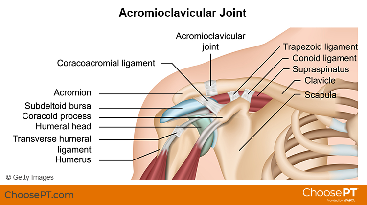
The acromioclavicular, or AC, joint is part of the shoulder girdle (the collar bone and shoulder blade that support the shoulder joint). An AC joint injury describes an injury to the top of the shoulder. It occurs where the front of the shoulder blade (acromion) attaches to the collarbone (clavicle). Most often, trauma, such as a fall directly on the outside of the shoulder, causes an AC joint injury. Overuse (repeated lifting of heavy weights or objects overhead with poor mechanics) also can result in an AC joint injury.

AC joint injuries are most common in people younger than age 35. Males sustain five times more traumatic AC joint injuries than females. Younger athletes who take part in activities like football, biking, skiing, and hockey have the highest risk for this injury.

Physical therapists can identify and effectively treat AC joint injuries, often avoiding the need for surgery.


What Are Acromicioclavicular (AC) Joint Injuries?

Acromioclavicular Joint Injury_ChoosePT_frame.png

There are four ligaments that hold the two bones of the AC joint together. When an AC joint injury occurs, these ligaments are stressed. This stress results in some degree of joint separation. There are two types of injuries of the AC joint: traumatic and overuse injuries.

Traumatic AC joint injury. This type of injury occurs when the joint is disrupted. The ligaments that hold the two bones of the joint together get stretched too far. This is called a shoulder separation. It is different from a shoulder dislocation, which involves the ball-and-socket joint.

Traumatic AC joint injuries are most common in people who fall and land on the outside of the shoulder or hand. Examples include a:

  • Football player who is tackled.

  • Cyclist who crashes.

  • Worker who falls off a ladder.

Traumatic AC joint injuries can range from a mild to severe grade. Grading is based on the amount of joint separation involved. Mild cases can be treated by a physical therapist. More severe cases may require surgery followed by physical therapy.

Overuse AC joint injury. This type of injury occurs over time due to repeated and too much stress on the joint. Cartilage at the end of the acromion and clavicle bones protects the joint from daily wear and tear. Over time, the demand placed on this cartilage may be more than it can endure. The result is an overuse injury that can lead to major wearing of the cartilage and arthritis. Overuse AC joint injury is most common in people who do repeated tasks. Examples include:

  • Heavy weightlifting (bench and military presses).

  • Jobs that require physical work with the arms stretched over the head.

How Does It Feel?

With an AC joint injury, you may experience:

  • General shoulder pain and swelling.

  • Swelling and tenderness over the AC joint.

  • Loss of shoulder strength.

  • A visible bump above the shoulder.

  • Pain when lying on the involved side.

  • Loss of shoulder motion.

  • A “popping” sound or feeling that your shoulder “catches” with movement.

  • Discomfort with daily activities that stress the AC joint. Examples include lifting objects overhead, reaching across your body, or carrying heavy objects at your side.

How Is It Diagnosed?

Physical therapists can diagnose an AC joint injury through a shoulder exam. Your physical therapist will conduct a full evaluation to find out the degree of your injury and identify all the factors that may contribute to it.

They will begin by interviewing you to learn about your health history. They may be helped by forms you fill out before your first session. The interview will become more specific to the condition of your shoulder. Your physical therapist may ask you questions such as:

  • How did your injury occur?

  • How have you taken care of the condition, such as seeing other health care providers? Have you had imaging (X-ray, MRI) or other tests and received their results?

  • What are your current symptoms? How have they changed your typical day and activities?

  • Do you have pain, and if so, what is the location and intensity of your pain? Does the pain vary during the day?

  • Do you have trouble doing any activities? What activities are you unable to do?

This information allows the physical therapist to better understand what you are going through. It also helps determine the course of your physical exam.

The physical exam will vary depending on your interview. Most often it will begin with observing the region of your symptoms and any movements or positions that cause pain. Your physical therapist also may examine other areas of your body that may have changed due to problems with your shoulder function. They may:

  • Watch you move your arm and shoulder overhead, and while doing other reaching tasks.

  • Assess the mobility and strength of your shoulder.

  • Check other regions of the body as needed. This will help to determine if other areas also require treatment to improve your condition.

  • Gently, but skillfully, feel around your shoulder and the AC joint to find exactly where it is most painful.

Your physical therapist will discuss their findings with you. They will work with you to develop a program for your specific needs and goals and to help you heal.

In some cases, your physical therapist may refer you for diagnostic imaging. Ultrasound, X-ray, or MRI can help to confirm the diagnosis and find out how severe the injury is.

How Can a Physical Therapist Help?

Your physical therapist will design a treatment plan to help you safely return to your desired activities. Your treatment plan may include:

  • Patient education. Your physical therapist will educate you about your AC joint and shoulder injury. They will work with you to identify any external factors causing your pain, including the amount and type of exercises and activities you do. Your physical therapist will recommend improvements to your activities.

  • Pain management. Your physical therapist will address your pain. This may include applying ice to the affected area and other methods. They also may recommend changing some activities that cause pain. Physical therapists are experts in prescribing pain-management methods. These can reduce or eliminate the need for medicines, including opioids.

  • Range-of-motion exercise. The mobility of the AC joint and shoulder may be limited, causing increased stress on the shoulder. Your physical therapist may teach you self-stretching methods. These can decrease tension and help restore normal motion of your injured joints.

  • Manual therapy. Your physical therapist may apply hands-on treatments to gently move your muscles and joints. These techniques help to improve movement. Your physical therapist also may use manual therapy to guide your shoulder area into a less stressful movement pattern.

  • Muscle strength. Muscle weaknesses or imbalances can contribute to problems of the AC joint and shoulder. They also can cause continued symptoms. Based on the how serious your injury is, your physical therapist will design a safe resistance program to aid your recovery. Exercises may include using resistance machines in the clinic and doing exercises to strengthen your core (midsection). You may begin doing exercises while lying on a table or at home on the bed or floor. You then may advance to exercises done in a standing position. Your physical therapist will choose what exercises are right for you based on your diagnosis, age, and condition. They will determine when it is safe for you to exercise on your own at home or in a gym.

  • Functional training. Once your pain, strength, and motion improve, functional training can help you safely resume more demanding activities. To minimize the stress to the AC joint and shoulder it is important to teach your body safe, controlled movements. Your physical therapist will create a series of activities to help you learn how to use and move your body correctly and safely. These may include retraining your movements and positioning when throwing, swinging a racket, lifting objects overhead, or doing other daily activities.

Can This Injury or Condition Be Prevented?

Accidents happen and it can be difficult to prevent many traumatic AC joint injuries. However, much can be done to prevent the string of events that lead to overuse injuries. Physical therapists can help reduce overuse injuries by:

  • Teaching you how to properly lift objects overhead at work.

  • Demonstrating good form for overhead resistance training or sports activities.

  • Helping you maintain general shoulder strength and motion to safely perform tasks.

Consult a physical therapist as soon as possible if you have persistent constant or worsening symptoms.

What Kind of Physical Therapist Do I Need?

All physical therapists are prepared through education and clinical experience to treat a variety of conditions or injuries. You may want to consider:

  • A physical therapist who is experienced in treating people with orthopedic or musculoskeletal (muscle, bone, and joint injuries).

  • A physical therapist who is a board-certified specialist or who has completed a residency in orthopedic or sports physical therapy. This physical therapist will have advanced knowledge, experience, and skills that apply to those who are physically active.

You can find physical therapists who have these and other credentials by using Find a PT, the online tool built by the American Physical Therapy Association. This tool can help you search for physical therapists with specific clinical expertise in your area.

General tips when you're looking for a physical therapist (or any other health care provider):

  • Get recommendations from family, friends, or other health care providers.

  • When you contact a physical therapy clinic for an appointment, ask about the physical therapists' experience in helping patients with shoulder pain.

  • During your first visit with the physical therapist, be prepared to describe your symptoms in as much detail as possible, and report activities and movements that make your symptoms worse.

Further Reading

The American Physical Therapy Association believes that consumers should have access to information to help them make informed health care decisions and prepare them for their visit with a health care provider.

The following resources offer some of the best scientific evidence related to physical therapy treatment for AC joint injuries. They report recent research and give an overview of the standards of practice both in the United States and internationally. They link to a PubMed* abstract that also may offer free access to the full text, or to other resources. You can read them or print out a copy to bring with you to your health care provider.

Frank RM, Cotter EJ, Leroux TS, Romeo AA. Acromioclavicular joint injuries: evidence-based treatment. J Am Acad Orthop Surg. 2019;27(17):e775–e788. Article Summary in PubMed.

Li X, Ma R, Bedi A, Dines DM, Altchek DW, Dines JS. Management of acromioclavicular joint injuries. J Bone Joint Surg Am. 2014;96(1):73–84. Article Summary in PubMed .

Harris KD, Deyle GD, Gill NW, Howes RR. Manual physical therapy for injection-confirmed nonacute acromioclavicular joint pain. J Orthop Sports Phys Ther. 2012;42(2):66–80. Article Summary in PubMed.

Pallis M, Cameron KL, Svoboda SJ, Owens BD. Epidemiology of acromioclavicular joint injury in young athletes. Am J Sports Med. 2012;40(9):2072–2077. Article Summary in PubMed .

*PubMed is a free online resource developed by the National Center for Biotechnology Information. PubMed contains millions of citations to biomedical literature, including citations in the National Library of Medicine's MEDLINE database.


Reviewed and revised in 2021 by Erin Hayden, PT, DPT, board-certified clinical specialist in orthopedic physical therapy, and Stephen Reischl, PT, DPT, board-certified clinical specialist in orthopedic physical therapy, on behalf of the Academy of Orthopaedic Physical Therapy. Authored in 2014 by Allison Mumbleau, PT, DPT, board-certified clinical specialist in sports physical therapy.


Patellofemoral Knee Pain

Patellofemoral pain syndrome (PFPS) refers to pain at the front of the knee, in and around the kneecap (patella). PFPS is one of the most common types of knee pain experienced in the United States, particularly among athletes, active teenagers, older adults, and people who perform physical labor. Patellofemoral pain affects more women than men and accounts for 20% to 25% of all reported knee pain. Physical therapists design exercise and treatment programs for people experiencing PFPS to help them reduce their pain, restore normal movement, and avoid future injury.

Current research indicates that PFPS is an "overuse syndrome," which means that it may result from repetitive or excessive use of the knee. Other contributing factors may include:

  • Weakness, tightness, or stiffness in the muscles around the knee and hip

  • An abnormality in the way the lower leg lines up with the hip, knee, and foot

  • Improper tracking of the kneecap

These conditions can interfere with the ability of the kneecap to glide smoothly on the femur (the bone that connects the knee to the thigh) in the femoral groove (situated along the thigh bone) during movement. The friction between the undersurface of the kneecap and the femur causes the pain and irritation commonly seen in PFPS. The kneecap also may fail to track properly in the femoral groove when the quadriceps muscle on the inside front of the thigh is weak, and the hip muscles on the outside of the thigh are tight. The kneecap gets pulled in the direction of the tight hip muscles and can track or tilt to the side, which irritates the tissues around the kneecap.

PFPS often occurs in people who are physically active or who have suddenly increased their level of activity, especially when that activity involves repeated knee motion, such as running, stair climbing, squatting, or repeated carrying of heavy loads. Older adults may experience age-related changes that cause the cartilage on the undersurface of the kneecap to wear out, resulting in pain and difficulty completing daily tasks without pain.

PatellofemoralPain_SM.jpg


How Does it Feel?

People with PFPS may experience:

  • Pain when walking up or down stairs or hills

  • Pain when walking on uneven surfaces

  • Pain that increases with activity and improves with rest

  • Pain that develops after sitting for long periods of time with the knee bent

  • A "crack" or "pop" when bending or straightening the knee

How Is It Diagnosed?

Your physical therapist will review your health history, perform a thorough examination, and conduct a series of tests to evaluate the knee. Your therapist may observe the alignment of your feet, analyze your walking and running patterns, and test the strength of your hip and thigh muscles to find out whether there is a weakness or imbalance that might be contributing to your pain. Your physical therapist also will check the flexibility of the muscles in your leg, paying close attention to those that attach at the knee.

Generally, X-rays are not needed to diagnose PFPS. Your physical therapist may consult with an orthopedic physician who may order an X-ray to rule out other conditions.

How Can a Physical Therapist Help?

After a comprehensive evaluation, your physical therapist will analyze the findings and, if PFPS is present, your therapist will prescribe an exercise and rehabilitation program just for you. Your program may include:

Strengthening exercises. Your physical therapist will teach you exercises targeted at the hip (specifically, the muscles of the buttock and thigh), the knee (specifically, the quadriceps muscle located on the front of your thigh that straightens your knee), and the ankle. Strengthening these muscles will help relieve pressure on the knee, as you perform your daily activities.

Stretching exercises. Your physical therapist also will choose exercises to gently stretch the muscles of the hip, knee, and ankle. Increasing the flexibility of these muscles will help reduce any abnormal forces on the knee and kneecap.

Positional training. Based on your activity level, your physical therapist may teach you proper form and positioning when performing activities, such as rising from a chair to a standing position, stair climbing, squatting, or lunging, to minimize excessive forces on the kneecap. This type of training is particularly effective for athletes.

Cross-training guidance. PFPS is often caused by overuse and repetitive activities. Athletes and active individuals can benefit from a physical therapist’s guidance about proper cross-training techniques to minimize stress on the knees.

Taping or bracing. Your physical therapist may choose to tape the kneecap to reduce your pain and retrain your muscles to work efficiently. There are many forms of knee taping, including some types of tape that help align the kneecap and some that just provide mild support to irritated tissues around it. In some cases, a brace may be required to hold the knee in the best position to ensure proper healing.

Electrical stimulation. Your physical therapist may prescribe treatments with gentle electrical stimulation to reduce pain and support the healing process.

Activity-based exercises. If you are having difficulty performing specific daily activities, or are an athlete who wants to return to a specific sport, your physical therapist will design individualized exercises to rebuild your strength and performance levels.

Fitting for an orthosis. If the alignment and position of your foot and arch appear to be contributing to your knee pain, your physical therapist may fit you with a special shoe insert called an orthosis. The orthosis can decrease the stress to your knee caused by low or high arches.

Can this Injury or Condition be Prevented?

PFPS is much easier to treat if it is caught early. Timely treatment by a physical therapist may help stop any underlying problems before they become worse. If you are experiencing knee pain, contact a physical therapist immediately. 

Your physical therapist can show you how to adjust your daily activities to safeguard your knees, and teach you exercises to do at home to strengthen your muscles and bones—and help prevent PFPS.

Physical therapists can assess athletic footwear and recommend proper choices for runners and daily walkers alike. Wearing the correct type of shoes for your activity and changing them when they are no longer supportive is essential to injury prevention.

Real Life Experiences

Amelia is a 25-year-old office assistant who loves to start her day with a 5-mile run. Over the past 6 months, she has been training for her first marathon. She began by training on very flat ground and has just moved to a hilly area.

Last week, Amelia began feeling pain in the front of her left knee when running downhill. Today, she had to stop running after 3 miles because of her knee pain. She called her physical therapist.

Amelia's physical therapist completes a comprehensive evaluation, including a screening for other possible conditions that might be causing her pain. He uses special tests to measure her strength and finds that she has weak hip muscles and tenderness around the kneecap. He determines that she has developed PFPS. Amelia is shocked to learn that she also has flat feet, and she’s not wearing the right supportive running shoes.

To begin her treatments, Amelia’s physical therapist applies special tape to the front of her knee to help reduce her pain, and instructs her in the use of ice to decrease her symptoms. He performs gentle movements of her kneecap and the surrounding tissues to help increase mobility and decrease pain. He teaches her special exercises to gently strengthen the weak muscles that support the knee.

He also designs a specific home-exercise program for Amelia to perform between sessions. He provides information about proper shoe choices for her foot and body type, and advises her to purchase shoes that will give her feet the right type of support. He also recommends that she try deep-water running or swimming for a week instead of her regular running program, until her condition improves.

After her first week of physical therapy, Amelia notices a decrease in her pain and an increased ability to walk up and down stairs without pain. Her physical therapist approves her new footwear, and adds more challenging exercises to her session and her home program. He gives her the go-ahead to race-walk. She applies ice only when she has pain.

After 2 weeks, Amelia reports she is feeling even less pain. Her physical therapist continues to increase the intensity of her exercises, and she starts to run again—but only on flat surfaces and short distances combined with longer walk intervals.

After a few more weeks of therapy, Amelia occasionally feels only slight twinges of pain and gradually resumes her prior level of training. Her physical therapist recommends continuation of her stretching and strengthening exercises, and discharges her from physical therapy.

A few months later, Amelia completes her first marathon pain free. She is thrilled to learn that her time was a personal best!

This story was based on a real-life case. Your case may be different. Your physical therapist will tailor a treatment program to your specific case.

What Kind of Physical Therapist Do I Need?

All physical therapists are prepared through education and experience to treat a variety of conditions or injuries. You may want to consider:

  • A physical therapist who is experienced in treating people with orthopedic, or musculoskeletal, problems.

  • A physical therapist who is a board-certified clinical specialist or who has completed a residency or fellowship in orthopedic physical therapy and has advanced knowledge, experience, and skills that may apply to your condition.

You can find physical therapists who have these and other credentials by using Find a PT, the online tool built by the American Physical Therapy Association to help you search for physical therapists with specific clinical expertise in your geographic area.

General tips when you're looking for a physical therapist:

  • Get recommendations from family and friends or from other health care providers.

  • When you contact a physical therapy clinic for an appointment, ask about the physical therapist's experience in helping people with patellofemoral pain syndrome.

During your first visit with the physical therapist, be prepared to describe your symptoms in as much detail as possible, and say what makes your symptoms worse.

Further Reading

The American Physical Therapy Association (APTA) believes that consumers should have access to information that could help them make health care decisions and also prepare them for their visit with their health care provider.

The following articles provide some of the best scientific evidence related to physical therapy treatment of patellofemoral pain syndrome. The articles report recent research and give an overview of the standards of practice both in the United States and internationally. The article titles are linked either to a PubMed* abstract of the article or to free access of the full article, so that you can read it or print out a copy to bring with you to your health care provider.

Tevhen DS, Robertson J. Knee pain: strengthen my hips? But it's my knees that hurt! J Orthop Sports Phys Ther. 2011-41-571. Article Summary on PubMed.

Davis IS, Powers CM. Patellofemoral pain syndrome: proximal, distal and local factors, an international retreat, April 30-May 2, 2009, Fells Point, Baltimore, MD. J Orthop Sports Phys Ther. 2010;40:A1–A16. Article Summary on PubMed.

Fukuda TY, Rossetto FM, Magalhaes E, et al. Short-term effects of hip abductors and lateral rotators strengthening in females with patellofemoral pain syndrome: a randomized controlled clinical trial. J Orthop Sports Phys Ther. 2010;40:736–742. Article Summary on PubMed.

Dixit S, DiFiori JP, Burton M, Mines B. Management of patellofemoral pain syndrome. Am Fam Physician. 2007;75:194–202. Free Article.

Powers CM, Ward SR, Chan LD, et al. The effect of bracing on patella alignment and patellofemoral joint contact area. Med Sci Sports Exerc. 2004;36:1226-1232. Article Summary on PubMed.

Bizzini M, Childs JD, Piva SR, Delitto A. Systematic review of the quality of randomized controlled trials for patellofemoral pain syndrome. J Ortho Sports Phys Ther. 2003;33:4–20. Article Summary on PubMed.

Crossley K, Bennell K, Green S, et al. Physical therapy for patellofemoral pain: a randomized, double-blinded, placebo-controlled trial. Am J Sports Med. 2002;30:857–865. Article Summary on PubMed.


* PubMed is a free online resource developed by the National Center for Biotechnology Information (NCBI).  PubMed contains millions of citations to biomedical literature, including citations in the National Library of Medicine’s MEDLINE database.

 Authored by Christopher Bise, PT, MS, DPT. Revised by Julie Mulcahy, PT. Reviewed by the MoveForwardPT.com editorial board.


Physical Therapist's Guide to Iliotibial Band Syndrome (ITBS or "IT Band Syndrome")

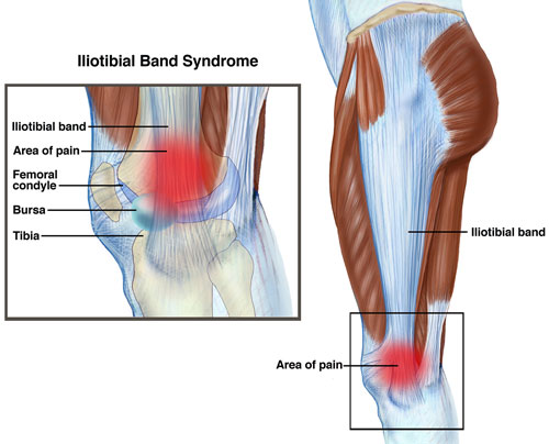
IliotibialBand_Small.jpg

Iliotibial band syndrome (ITBS) is one of the most common causes of knee pain, particularly in individuals involved in endurance sports. It accounts for up to 12% of running injuries and up to 24% of cycling injuries. ITBS is typically managed conservatively through physical therapy and temporary activity modification.

What is Iliotibial Band Syndrome (ITBS)?

Iliotibial band syndrome (ITBS) occurs when excessive irritation causes pain at the outside (or lateral) part of the knee. The iliotibial band (ITB), often referred to as the "IT band" is a type of soft tissue that runs along the side of the thigh from the pelvis to the knee. As it approaches the knee, its shape thickens as it crosses a prominent area of the thigh (femur) bone, called the lateral femoral condyle. Near the pelvis, it attaches to 2 important hip muscles, the tensor fascia latae (TFL) and the gluteus maximus.

Irritation and inflammation arise from friction between the ITB and underlying structures when an individual moves through repetitive straightening (extension) and bending (flexion) of the knee. Typically, ITBS pain occurs with overuse during activities such as running and cycling.

ITBS involves many lower extremity structures, including muscles, bones, and other soft tissues. Usually discomfort arises from:

  • Abnormal contact between the ITB and thigh (femur) bone
  • Poor alignment and/or muscular control of the lower body
  • Prolonged pinching (compression) or rubbing (shearing) forces during repetitive activities

The common structures involved in ITBS are:

  • Iliotibial band
  • Bursa (fluid-filled sack that sits between bones and soft tissues to limit friction)
  • Hip muscles

ITBS can occur in:

  • Athletes performing repetitive activities, such as squatting, and endurance sports such as running and cycling
  • Individuals who spend long periods of time in prolonged positions, such as sitting or standing for a long workday, climbing or squatting, or kneeling
  • Individuals who quickly start a new exercise regimen without proper warm-up or preparation

Back to Top


Signs and Symptoms

With ITBS, you may experience:

  • Stabbing or stinging pain along the outside of the knee
  • A feeling of the ITB “snapping” over the knee as it bends and straightens
  • Swelling near the outside of your knee
  • Occasionally, tightness and pain at the outside of the hip
  • Continuous pain following activity, particularly with walking, climbing, or descending stairs, or moving from a sitting to standing position

Pain is usually most intense when the knee is in a slightly bent position, either right before or right after the foot strikes the ground. This is the point where the ITB rubs the most over the femur.

Back to Top


How Is It Diagnosed?

Your physical therapist will ask you questions about your medical history and activity regimen. A physical examination will be performed so that your physical therapist can collect movement (range of motion), strength, and flexibility measurements at the hip, knee, and ankle.

When dealing with ITBS, it is also common for a physical therapist to use special tests and complete a movement analysis, which will provide information on the way that you move and how it might contribute to your injury. This could include assessment of walking/running mechanics, foot structure, and balance. Your therapist may have you repeat the activity that causes your pain to see firsthand how your body moves when you feel pain. If you are an athlete, your therapist might also ask you about your chosen sport, shoes, training routes, and exercise routine.

Typically, medical imaging tests, such as x-ray and MRI, are not needed to diagnosis ITBS.

Back to Top


How Can a Physical Therapist Help?

Your physical therapist will use treatment strategies to focus on:

Range of motion

Often, abnormal motion of the hip and knee and foot joint can cause ITBS because of how the band attaches to hip muscles. Your therapist will assess the motion of your injury leg compared with expected normal motion and the motion of the hip on your uninvolved leg.

Muscle strength

Hip and core weakness can contribute to ITBS. The "core" refers to the muscles of the abdomen, low back, and pelvis. Core strength is important, as a strong midsection will allow greater stability through the body as the arms and legs go through various motions. For athletes performing endurance sports, it is important to have a strong core to stabilize the hip and knee joints during repetitive leg motions. Your physical therapist will be able to determine which muscles are weak and provide specific exercises to target these areas.

Manual therapy

Many physical therapists are trained in manual therapy, which means they use their hands to move and manipulate muscles and joints to improve motion and strength. These techniques can target areas that are difficult to treat on your own.

Functional training

Even when an individual has normal motion and strength, it is important to teach the body how to perform controlled and coordinated movements so there is no longer excessive stress at the previously injured structures. Your physical therapist will develop a functional training program specific to your desired activity. This means creating exercises that will replicate your activities and challenge your body to learn the correct way to move.

Your physical therapist will also work with you to develop an individualized treatment program specific to your personal goals. He or she will offer tips to help you prevent your injury from reoccurring.

Back to Top


Can this Injury or Condition be Prevented?

Maintaining core and lower extremity strength and flexibility and monitoring your activity best prevents ITBS. It is important to modify your activity and contact your physical therapist soon after first feeling pain. Research indicates that when soft tissues are irritated and the offending activity is continued, the body does not have time to repair the injured area. This often leads to persistent pain, and the condition becomes more difficult to resolve.

Once you are involved in a rehabilitation program, your physical therapist will help you determine when you are ready to progress back to your previous activity level. He or she will make sure that your body is ready to handle the demands of your activities so that your injury does not return. You will also receive a program to perform at home that will help you maintain the improvements that you gained during rehabilitation.

Back to Top


Real Life Experiences

Sarah is a 31-year-old mother training for her first triathlon. With a young child at home, she has to squeeze in her training sessions early in the morning. She rarely has time to cool down or stretch after riding her bike or running because she has to get home before her child wakes up.

Sarah signs up for her first race and begins to increase her cycling and running in preparation. One day during the middle of a long run, she feels a sharp pain at the outside of her knee. It starts hurting with every step, and doesn't go away, even after she stops and stretches. Far from home, she has to finish her run despite the nagging pain. When she gets home, she puts ice on it, but for the rest of the day she has trouble going up and down stairs, or squatting to pick up her son, and feels pain when standing up after driving the car. The next day, she tries to ride her bike, but the knee pain is still there and feels worse.

Wisely, Sarah stops running and cycling and contacts her physical therapist.

Sarah's physical therapist conducts a comprehensive evaluation of her hip and knee motion, strength, balance, and running mechanics. She uses special tests and measures to determine if Sarah’s pain is related to her iliotibial band or if there are other problems occurring simultaneously. She talks with Sarah about her training routine, including equipment (shoes, position on the bike, etc), the routes she runs and their surfaces, and her stretching program. The therapist diagnoses Sarah with iliotibial band syndrome. She guides Sarah through specific exercises in the clinic, including manual stretching of the hip joint by the therapist, sidelying leg raises for hip strengthening, and single leg squats to promote integrated core, hip, knee, and ankle function. Sarah will also perform these exercises at home as a part of her daily exercise routine to maximize improvement and help ensure her sustainable success.

Sarah's physical therapist helps her develop strategies for training, taking into consideration her lifestyle as a busy mother, to help her stay injury-free. Together, they outline a 6-week rehabilitation program for iliotibial band syndrome. Sarah will come to the clinic 1-2 times each week, where her therapist will assess her progress, perform manual therapy techniques, and advance her exercise program as appropriate. Sarah will also have a daily exercise routine to perform independently at home, including stretching and strengthening activities.

In 6 weeks, Sarah has met all of her physical therapy goals and completes her rehabilitation in the clinic. After building her training gradually over the next month, she is able to train and successfully crosses the finish line just as planned!

Back to Top


What Kind of Physical Therapist Do I Need?

All physical therapists are prepared through education and clinical experience to treat a variety of conditions or injuries. You may want to consider:

  • A physical therapist who is experienced in treating people with orthopedic, or musculoskeletal, injuries.
  • A physical therapist who is a board-certified specialist or who has completed a residency in orthopedic or sports physical therapy, as he or she will have advanced knowledge, experience, and skills that apply to an athletic population.

You can find physical therapists that have these and other credentials by using Find a PT, the online tool built by the American Physical Therapy Association to help you search for physical therapists with specific clinical expertise in your geographic area.

General tips when you're looking for a physical therapist (or any other health care provider):

  • Get recommendations from family and friends or from other health care providers.
  • When you contact a physical therapy clinic for an appointment, ask about the physical therapists' experience in helping people with ITBS.
  • During your first visit with the physical therapist, be prepared to describe your symptoms in as much detail as possible, and report activities that make your symptoms worse.

Back to Top


Further Reading

The American Physical Therapy Association believes that consumers should have access to information that could help them make health care decisions and also prepare them for their visit with their health care provider.

The following articles provide some of the best scientific evidence related to physical therapy treatment of ITBS. The articles report recent research and give an overview of the standards of practice both in the United States and internationally. The article titles are linked either to a PubMed* abstract of the article or to free full text, so that you can read it or print out a copy to bring with you to your health care provider.

Strauss EJ, Kim S, Calcei JG, Park D. Iliotibial band syndrome: evaluation and management. J Am Acad Orthop Surg. 2011;19:726–736. Free Article.

Ellis R, Hing W, Reid D. Iliotibial band friction syndrome: a systematic review. Man Ther. 2007;12:200–208. Article Summary on PubMed.

Fredericson M, Weir A. Practical management of iliotibial band syndrome in runners. Clin J Sports Med. 2006;16:261–268. Article Summary on PubMed.

Fredericson M, Wolf C. Iliotibial band syndrome in runners: innovations in treatment. Sports Med. 2005;35:451–459. Article Summary on PubMed.

Levin J. Run down: battling IT band syndrome in long distance runners. Biomechanics. 2003;1:22–25. Article Summary Not Available.

Fredericson M, Cookingham CL, Chaudhari AM, et al. Hip abductor weakness in distance runners with iliotibial band syndrome. Clin J Sports Med. 2000;10:169–175. Article Summary on PubMed.

* PubMed is a free online resource developed by the National Center for Biotechnology Information (NCBI). PubMed contains millions of citations to biomedical literature, including citations in the National Library of Medicine’s MEDLINE database.

Authored by Laura Stanley, PT, DPT, SCS. Reviewed by the MoveForwardPT.com editorial board.