Physical Therapist's Guide to Patellofemoral Pain

What Is Patellofemoral Pain?

PFP may occur after a sudden increase in activities like running or jumping. Research suggests that PFP results from activity levels that are increased faster than the knee can adapt. Other contributing factors to PFP may include:

  • Weakness of the thigh muscles.

  • Specializing in a single sport, which requires repeating the same movements again and again.

  • Certain hip and knee coordination patterns during running and jumping activities.

PFP does not go away on its own. If you have symptoms of PFP, it’s important to seek care from a physical therapist so you can return to the activities that you enjoy.

How Does it Feel?

People with PFP may experience pain:

  • When walking up or down stairs or hills.

  • When playing a sport.

  • With deep knee bending (squatting).

  • When walking on uneven surfaces.

  • With activity, but improving with rest.

  • After sitting for long periods of time with the knee bent.

How Is It Diagnosed?

APTA-Knee-Joint-Patella-Pain_750x419.jpg

Your physical therapist will review your health history and conduct a series of tests to evaluate you and your knee. PFP is diagnosed by analyzing any movement that causes pain, and ruling out other possible conditions.

Your physical therapist may analyze your walking and running patterns. They may test the strength of your hip and thigh muscles to find out if weakness is contributing to your pain. Medical imaging, such as an X-ray or MRI, is not helpful in diagnosing PFP. However, your physical therapist may consult with an orthopedic physician who may order imaging to rule out other conditions.

How Can a Physical Therapist Help?

If PFP is diagnosed, your physical therapist will develop an exercise and rehabilitation program just for you. Your program may include:

Strengthening exercises. Your physical therapist will teach you exercises to help strengthen the muscles around the hip and the knee itself. Research shows that this type of exercise therapy is the best approach to managing PFP.

Taping. Your physical therapist may teach you how to apply tape to your knee, which may improve your ability to perform exercises that would normally be painful. However, taping alone will not resolve PFP. It must only be used along with your exercise program.

Shoe inserts. Your physical therapist may recommend shoe inserts to help reduce your pain when exercising. But inserts alone, like taping, will not treat PFP. Your physical therapist will design an exercise program to fit your specific needs and goals.

Coordination training. Based on your activity level, your physical therapist may help retrain your hip and knee movement patterns to reduce your knee pain.

This type of training is effective for athletes, in particular, and may focus on movements like:

  • Stair climbing.

  • Squatting.

  • Running and jumping.

Cross-training guidance. Physical therapists help athletes and active people perform different movements (cross-training). This helps them stay active until they can return to a favorite activity.

Return to full activity. Your physical therapist will help guide a gradual return to your favorite activities, such as running and jumping, and will teach you good overall exercise habits to help maximize the health of your knee.

Treatments That Do Not Work for PFP

While these can be appropriate for the treatment of other injuries or conditions, the following do notwork for PFP:

Quick fixes. “Passive” treatments like dry needling, ultrasound, laser, or electrical stimulation are not helpful for people with PFP. The most effective treatment for PFP is an exercise program that targets the hip and knee muscles.

Rest. If you are experiencing PFP, it is important to understand that rest only helps temporarily. Your pain will likely return when you go back to your normal activity. Rest is not helpful in the long term. A movement program guided by your physical therapist is your best treatment option.

Can this Injury or Condition be Prevented?

Current research shows that a person’s age, height, body weight, or foot alignment may not contribute to the risk of developing PFP at all. A knock-kneed posture also does not increase the risk of developing PFP.

However, a few preventive measures can be effective. To help reduce your risk of developing PFP:

  • Keep your thigh muscles strong.

  • Maintain good exercise habits.

  • Avoid rapid spikes in activity levels.

  • Participate in a variety of sports, rather than just repeating the same movements again and again.

What Kind of Physical Therapist Do I Need?

All physical therapists are prepared through education and experience to treat a variety of conditions or injuries including PFP. However, you may want to consider:

  • A physical therapist who is experienced in treating people with orthopedic, or musculoskeletal, problems.

  • A physical therapist who is a board-certified clinical specialist or who has completed a residency or fellowship in orthopedic physical therapy. This physical therapist will have advanced knowledge, experience, and skills that may apply to your condition.

  • While it may be tempting to seek quick fixes for your knee pain, there is no evidence that passive treatments work for persons with PFP. If you have PFP, seek care from a physical therapist who uses progressive exercise therapy for the treatment of this condition.

You can find physical therapists who have these and other credentials by using Find a PT, an online tool provided by the American Physical Therapy Association. You can search for physical therapists with specific clinical expertise in your geographic area.

General tips when you're looking for a physical therapist:

  • Get recommendations from family and friends or from other health care providers.

  • When you contact a physical therapy clinic for an appointment, ask about the physical therapist's experience in helping people with patellofemoral pain (PFP).

  • During your first visit with a physical therapist, you will be asked to describe your symptoms in as much detail as possible, and say what makes your symptoms worse. Here are some tips to prepare for your visit.

Further Reading

The American Physical Therapy Association (APTA) believes that consumers should have access to information that could help them make health care decisions and prepare for a visit with their health care provider.

The following articles provide some of the best scientific evidence related to physical therapy treatment of patellofemoral pain syndrome. The articles report recent research and give an overview of the standards of practice both in the United States and internationally. The article titles are linked either to a PubMed* abstract of the article or to free access of the full article, so you can read it or print out a copy to bring with you to your health care provider.

Patellofemoral pain: treating painful kneecaps. J Orthop Sports Phys Ther. 2019;49(9):633. Free Article.

Willy RW, Hoglund LT, Barton CJ, et al. Patellofemoral Pain. J Orthop Sports Phys Ther.2019;49(9):CPG1–CPG95. Free Article.

Neal BS, Lack SD, Lankhorst NE, Raye A, Morrissey D, van Middelkoop M. Risk factors for patellofemoral pain: a systematic review and meta-analysis. Br J Sports Med. 2019;53(5):270–281. Free Article.

Lack S, Barton C, Sohan O, Crossley K, Morrissey D. Proximal muscle rehabilitation is effective for patellofemoral pain: a systematic review with meta-analysis. Br J Sports Med. 2019;49(21):1365-1376. Free Article.

* PubMed is a free online resource developed by the National Center for Biotechnology Information (NCBI). PubMed contains millions of citations to biomedical literature, including citations in the National Library of Medicine’s MEDLINE database.

Authored by Christopher Bise, PT, DPT, MS. Revised and reviewed by Richard Willy, PT, PhD.

Pes Anserine Bursitis

Pes anserine bursitis is a condition that produces pain on the inside of the knee and lower leg. It occurs most commonly in young people involved in sports (eg, running or swimming the breaststroke), middle-aged women who are overweight, and people aged 50 to 80 years who have osteoarthritis of the knee. Up to 75% of people who have osteoarthritis of the knee have symptoms of pes anserine bursitis. The condition is also commonly associated with type 2 diabetes; 24% to 34% of patients with type 2 diabetes who report knee pain are found to have pes anserine bursitis. However, in some cases no direct cause can be identified. Physical therapists treat people with pes anserine bursitis to reduce pain, swelling, stiffness, and weakness, as well as identify and treat the underlying cause of the condition.

download.jpg

What is Pes Anserine Bursitis?

The pes anserine bursa is a small, fluid-filled sac located 2 to 3 inches below the knee joint on the inside of the lower leg. It lies beneath 3 tendons that attach to muscles of the thigh, and prevents the tendons from rubbing on the shinbone (tibia). The term “bursitis” describes a condition where the bursa has become irritated and inflamed. This condition is usually painful and associated with some swelling in the affected area. Certain positions, motions, or disease processes can cause increased friction or stress on the bursa, leading to the development of bursitis.

Pes anserine bursitis can be caused by:

  • Repetitive activities, like squatting, stair climbing, and other work or household activities that are often repeated

  • Incorrect sports training techniques, such as a lack of stretching, sudden increases in run distances, or too much uphill running

  • Obesity

  • Osteoarthritis of the knee

  • Valgus positioning of the knee (ie, a “knock-knee” position where the knees angle inward)

  • Turning the leg sharply with the foot planted on the ground

  • Injury, such as a direct hit to the leg

  • Tight hamstring (back of the thigh) muscles

  • A tear in the cartilage of the knee

  • Flat feet

How Does it Feel?

With pes anserine bursitis, you may experience:

  • Pain and swelling on the inside of the lower leg, 2 to 3 inches below the knee joint; this pain may also extend to the front of the knee and down the lower leg

  • Pain when touching the inside of the lower leg, 2 to 3 inches below the knee joint

  • Pain when bending or straightening the knee

  • Pain or difficulty walking, sitting down, rising from a chair, or climbing stairs

How Is It Diagnosed?

Your physical therapist will conduct a thorough examination that includes taking your health history as well as asking you detailed questions about your injury, such as:

  • How and when did you notice the pain?

  • Did you feel pain or hear a "pop" when you injured your leg?

  • Did you turn your leg with your foot planted on the ground?

  • Did you change direction quickly while running?

  • Did you receive a direct hit to the leg while your foot was planted on the ground?

  • Did you see swelling around the knee in the first 2 to 3 hours following the injury?

  • Does your knee feel like it is buckling or “giving way” when you try to use it?

Your physical therapist also will perform special tests to help determine the likelihood that you have pes anserine bursitis. Your therapist may:

  • Gently press on the inner side of your knee to see if it is painful to the touch

  • Assess the range of motion you have at the knee and hip, as well as the strength of some of the muscles at these joints

  • Observe how you are walking, squatting, and performing other functional and sports-specific tasks as appropriate

To provide a definitive diagnosis, your physical therapist may collaborate with an orthopedic physician or other health care provider, who may order further tests, such as an x-ray, to confirm the diagnosis and to rule out other damage to the knee.

How Can a Physical Therapist Help?

Your physical therapist will work with you to design a specific treatment program that will speed your recovery, including exercises and treatments that you can do at home. Physical therapy will help you return to your normal lifestyle and activities.

The First 24 to 48 Hours

If you see a physical therapist within 24 to 48 hours of your injury, your therapist may advise you to:

  • Rest the area by avoiding walking or any activity that causes pain.

  • Apply ice packs to the area for 15 to 20 minutes every 2 hours.

  • Consult with a physician for further services, such as medication or diagnostic tests.

Individualized Treatment

Depending on your condition and goals, your individualized rehabilitation plan may include treatments to:

Reduce pain and swelling. Your physical therapist may use different types of treatments to control and reduce your pain and swelling, including ice, heat, ultrasound, electrical stimulation, taping, exercises, and hands-on therapy, such as massage.

Improve motion. Your physical therapist will choose specific activities and treatments to help restore normal movement in the knee and leg. These might begin with "passive" motions that the physical therapist performs for you to gently move your leg and knee joint, and then progress to active exercises that you do yourself.

Improve flexibility. Pes anserine bursitis is often related to tight hamstring (back of the thigh) muscles. Your physical therapist will determine if your hamstring muscles or any other leg muscles are tight, and teach you how to stretch them.

Improve strength. Certain exercises will aid healing at each stage of recovery. Your physical therapist will choose and teach you the correct exercises and equipment to steadily restore your muscle strength and power. These may include using cuff weights, stretch bands, weight-lifting equipment, and cardio-exercise equipment, such as treadmills or stationary bicycles.

Improve balance. Regaining your sense of balance is important after an injury. Your physical therapist may teach you exercises to improve your balance skills.

Speed recovery time. Your physical therapist is trained and experienced in choosing the best treatments and exercises to help you heal, return to a normal lifestyle, and reach your goals faster than you are likely to do on your own.

Return to activities. Initially, your physical therapist may recommend that you reduce or eliminate activities that aggravate your condition for a period of time. Your physical therapist will discuss your goals with you and set up a treatment program to help you meet them in the safest, fastest, and most effective way possible. You may learn specific exercises, work retraining activities, and sport-specific techniques and drills to help you achieve your own unique goals.

Other Treatment Options

Studies have shown that some patients who do not respond to conservative treatment, such as physical therapy, may benefit from medical therapy. Your physical therapist may recommend that you discuss other treatment options with your physician, including surgery. Although surgery is rarely prescribed for pes anserine bursitis, it sometimes is needed. If surgery is required for your condition, you will follow a recovery program over several weeks guided by your physical therapist. Your physical therapist will help you minimize pain, regain motion and strength, and return to normal activities in the safest and speediest manner possible.

Can this Injury or Condition be Prevented?

Your physical therapist can recommend a home-exercise program to strengthen and stretch the muscles around your knees, upper legs, and abdomen to help prevent the onset or recurrence of pes anserine bursitis. These may include strength and flexibility exercises for the legs, knees, and core muscles.

To help prevent a recurrence of the injury, or prevent its onset if you seek guidance before injury, your physical therapist may advise you to:

  • Learn correct knee positioning when participating in athletic activities.

  • Follow a consistent flexibility and strength exercise program, especially for the leg and hip muscles, to maintain good physical conditioning.

  • Practice balance and agility exercises and drills.

  • Always warm up before starting a sport or heavy physical activity.

  • Avoid sudden increases in running mileage or uphill running.

  • Wear shoes that are in good condition and fit well.

  • Maintain a healthy weight.

  • Treat and manage diabetes very closely.

  • Wear orthotics to reduce flat feet and valgus (knock-knee) positioning of the lower extremities.

  • Wear a knee brace to support the knee and reduce strain on the inside of the joint.

Real Life Experiences

Martha is a 40-year-old secretary who has become obese. Her goal is to lose 100 pounds with diet and exercise. To reach that goal, Martha recently joined a gym, and decided to try the latest craze—a Zumba class. Martha enjoyed the first week of classes, but when leaving the gym after the fourth class, she felt a sharp pain in the inner, lower side of her right knee. It got worse when she bent and straightened her knee and when she walked upstairs to go to bed that night. The next day, the pain was still there, making it hard for her to get to work. She contacted her physical therapist.

Martha’s physical therapist performed special tests on the tendons and muscles around the knee, and found that her hamstring (back of the thigh) muscles were extremely tight and her quadriceps (front of the thigh) muscles were weak. Martha’s knee was tender to the touch, and mildly swollen 2 to 3 inches below the knee joint on the inner side of the leg, where the pes anserine bursa is located.

Martha's physical therapist explained that her pes anserine bursa was irritated and swollen. He applied ice and electrical stimulation to the area for 20 minutes. He also applied some tape to gently support Martha's hamstring muscles and alleviate the swelling and pain. He showed her how to stretch her hamstring muscles at home, and how to apply ice every few hours. He recommended that she not attend her Zumba class until her symptoms cleared up.

When Martha returned for her next visit, her physical therapist taught her some exercises to improve the strength of the muscles of her legs and “core,” and to improve her balance. Martha and her physical therapist worked together consistently over the next few weeks. Her treatment program, both in the clinic and at home, as well as her return to activity, were carefully adjusted to help ensure her safe and effective recovery.

Martha received physical therapy treatments for 6 weeks, at which time she felt almost 100% pain free—and much stronger. Martha returned to the gym to perform the exercises and stretches she learned in physical therapy as well as a modified fitness program. By the fourth week, she was able to participate in half of the Zumba class and by the fifth week, to finish the full class.

Martha has continued to do the stretches and exercises she learned from her physical therapist, and is proud to report to her friends and family that she is now pain free—and losing weight!

What Kind of Physical Therapist Do I Need?

All physical therapists are prepared through education and experience to treat pes anserine bursitis. However, you may want to consider:

  • A physical therapist who is experienced in treating people with orthopedic injuries. Some physical therapists have a practice with an orthopedic focus.

  • A physical therapist who is a board-certified clinical specialist or who has completed a residency or fellowship in orthopedic or sports physical therapy. This physical therapist has advanced knowledge, experience, and skills that may apply to your condition.

You can find physical therapists who have these and other credentials by using Find a PT, the online tool built by the American Physical Therapy Association to help you search for physical therapists with specific clinical expertise in your geographic area.

General tips when you're looking for a physical therapist (or any other health care provider):

  • Get recommendations from family and friends, or from other health care providers.

  • When you contact a physical therapy clinic for an appointment, ask about the physical therapists' experience in helping people who have your type of injury.

  • During your first visit with the physical therapist, be prepared to describe your symptoms in as much detail as possible, and say what makes your symptoms worse.

Further Reading

The American Physical Therapy Association (APTA) believes that consumers should have access to information that could help them make health care decisions and also prepare them for a visit with their health care provider.

The following articles provide some of the best scientific evidence related to physical therapy treatment of pes anserine bursitis. The articles report recent research and give an overview of the standards of practice both in the United States and internationally. The article titles are linked either to a PubMed* abstract of the article or to free full text, so that you can read it or print out a copy to bring with you to your health care provider.

Uysal F, Akbal A, Gökmen F, Adam G, Reşorlu M. Prevalence of pes anserine bursitis in symptomatic osteoarthritis patients: an ultrasonographic prospective study. Clin Rheumatol. 2015;34(3):529–533. Article Summary in PubMed.

Klontzas ME, Akoumianakis ID, Vagios I, Karantanas AH. MR imaging findings of medial tibial crest friction. Eur J Radiol. 2013;82(11):e703–e706. Article Summary in PubMed.

Chatra PS. Bursae around the knee joints. Indian J Radiol Imaging. 2012;22(1):27-30. Free Article.

Helfenstein M Jr, Kuromoto J. Anserine syndrome [article in English and Portuguese]. Rev Bras Reumatol. 2010;50(3):313–327. Free Article.

Alvarez-Nemegyei J. Risk factors for pes anserinus tendinitis/bursitis syndrome: a case control study. J Clin Rheumatol. 2007;13(2):63–65. Article Summary in PubMed

Rennie WJ, Saifuddin A. Pes anserine bursitis: incidence in symptomatic knees and clinical presentation. Skeletal Radiol. 2005;34:395–398. Article Summary in PubMed

Handy JR. Anserine bursitis: a brief review. South Med J. 1997;90(4):376–377. Article Summary on PubMed.

Butcher JD, Salzman KL, Lillegard WA. Lower extremity bursitis [published correction in: Am Fam Physician. 1996;54(2):468]. Am Fam Physician. 1996;53(7):2317-2324. Article Summary on PubMed.

Hemler DE, Ward WK, Karstetter KW, Bryant PM. Saphenous nerve entrapment caused by pes anserine bursitis mimicking stress fracture of the tibia. Arch Phys Med Rehabil. 1991;72(5):336–337. Article Summary on PubMed.

*PubMed is a free online resource developed by the National Center for Biotechnology Information (NCBI).  PubMed contains millions of citations to biomedical literature, including citations in the National Library of Medicine’s MEDLINE database.

Revised by Daniel Farwell, PT, DPT, a board-certified sports clinical specialist. Authored by Andrea Avruskin, PT. Reviewed by the editorial board.

Osteoarthritis of the Knee

Osteoarthritis of the knee (knee OA) is the inflammation and wearing away of the cartilage on the bones that form the knee joint (osteo=bone, arthro=joint, itis=inflammation). The diagnosis of knee OA is based on 2 primary findings: radiographic evidence of changes in bone health (through medical images such as X-ray and magnetic resonance imaging [MRI]), and an individual’s symptoms (how you feel). Approximately 14 million people in the United States have symptomatic knee OA. Although more common in older adults, 2 million of the 14 million people with symptomatic knee OA were younger than 45 when diagnosed, and more than half were younger than 65.

download.jpg

What is Osteoarthritis of Knee?

Knee osteoarthritis (knee OA) is a progressive disease caused by inflammation and degeneration of the knee joint that worsens over time. It affects the entire joint, including bone, cartilage, ligaments, and muscles. Its progression is influenced by age, body mass index (BMI), bone structure, genetics, muscular strength, and activity level. Knee OA also may develop as a secondary condition following a traumatic knee injury. Depending on the stage of the disease and whether there are associated injuries or conditions, knee OA can be managed with physical therapy. More severe or advanced cases may require surgery.


How Does it Feel?

Individuals who develop knee OA may experience a wide range of symptoms and limitations based on the progression of the disease. Pain occurs when the cartilage covering the bones of the knee joint wears down. Areas where the cartilage is worn down or damaged exposes the underlying bone. The exposure of the bone allows increased stress and compression to the cartilage, and at times bone-on-bone contact during movement, which can cause pain. Because the knee is a weight-bearing joint, your activity level, and the type and duration of your activities usually have a direct impact on your symptoms. Symptoms may be worse with weight-bearing activity, such as walking while carrying a heavy object.

Symptoms of knee OA may include:

  • Worsening pain during or following activity, particularly with walking, climbing, or descending stairs, or moving from a sitting to standing position

  • Pain or stiffness after sitting with the knee bent or straight for a prolonged period of time

  • A feeling of popping, cracking, or grinding when moving the knee

  • Swelling following activity

  • Tenderness to touch along the knee joint

Typically these symptoms do not occur suddenly or all at once, but instead develop gradually over time. Sometimes individuals do not recognize they have osteoarthritis because they cannot remember a specific time or injury that caused their symptoms. If you have had worsening knee pain for several months that is not responding to rest or a change in activity, it is best to seek the advice of a medical provider.


How Is It Diagnosed?

Knee OA is diagnosed by 2 primary processes. The first is based on your report of your symptoms and a clinical examination. Your physical therapist will ask you questions about your medical history and activity. The therapist will perform a physical exam to measure your knee's movement (range of motion), strength, mobility, and flexibility. You might also be asked to perform various movements to see if they increase or decrease the pain you are experiencing.

The second tool used to diagnose knee OA is diagnostic imaging. Your physical therapist may refer you to a physician, who will order X-rays of the knee in a variety of positions to check for damage to the bone and cartilage of your knee joint. If more severe joint damage is suspected, an MRI may be ordered to look more closely at the overall status of the joint and surrounding tissues. Blood tests also may be ordered to help rule out other conditions that can cause symptoms similar to knee OA.


How Can a Physical Therapist Help?

Once you have received a diagnosis of knee OA, your physical therapist will design an individualized treatment program specific to the exact nature of your condition and your goals. Your treatment program may include:

Range-of-motion exercises. Abnormal motion of the knee joint can lead to a worsening of OA symptoms when there is additional stress on the joint. Your physical therapist will assess your knee’s range of motion compared with expected normal motion and the motion of the knee on your uninvolved leg. Your range-of-motion exercises will focus on improving your ability to bend and straighten your knee, as well as improve your flexibility to allow for increased motion.

Muscle strengthening. Strengthening the muscles around your knee will be an essential part of your rehabilitation program. Individuals with knee OA who adhere to strengthening programs have been shown to have less pain and an improved overall quality of life. There are several factors that influence the health of a joint: the quality of the cartilage that lines the bones, the tissue within and around the joints, and the associated muscles. Due to the wear and tear on cartilage associated with knee OA, maintaining strength in the muscles near the joint is crucial to preserve joint health. For example, as the muscles along the front and back of your thigh (quadriceps and hamstrings) cross the knee joint, they help control the motion and forces that are applied to the bones.

Strengthening the hip and core muscles also can help balance the amount of force on the knee joint, particularly during walking or running. The “core” refers to the muscles of the abdomen, low back, and pelvis. A strong core will increase stability throughout your body as you move your arms and legs. Your physical therapist will assess these different muscle groups, compare the strength in each limb, and prescribe specific exercises to target your areas of weakness.

Manual therapy. Physical therapists are trained in manual (hands-on) therapy. Your physical therapist will gently move your muscles and joints to improve their motion, flexibility, and strength. These techniques can target areas that are difficult to treat on your own. The addition of manual therapy techniques to exercise plans has been shown to decrease pain and increase function in people with knee OA.

Bracing. Compressive sleeves placed around the knee may help reduce pain and swelling. Devices such as realignment and off-loading braces are used to modify the forces placed on the knee. These braces can help "unload" certain areas of your knee and move contact to less painful areas of the joint during weight-bearing activities. Depending on your symptoms and impairments, your physical therapist will help determine which brace may be best for you.

Activity recommendations. Physical therapists are trained to understand how to prescribe exercises to individuals with injuries or pain. Since knee OA is a progressive disease, it is important to develop a specific plan to perform enough activity to address the problem, while avoiding excessive stress on the knee joint. Activity must be prescribed and monitored based on the type, frequency, duration, and intensity of your condition, with adequate time allotted for rest and recovery. Research has shown that individuals with knee OA who walked more steps per day were less likely to develop functional problems in the future. Your physical therapist will consider the stage and extent of your knee OA and prescribe an individualized exercise program to address your needs and maximize the function of your knee.

Modalities. Your physical therapist may recommend therapeutic modalities, such as ice and heat, to aid in pain management.

If Surgery Is Required

The meniscus (the shock absorber of the knee) may be involved in some cases of knee OA. In the past, surgery (arthroscopy) to repair or remove parts or all of this cartilage was common. Current research, however, has shown that—in a group of patients who were deemed surgical candidates for knee OA with involvement of the meniscus—60% to 70% of those who first participated in a physical therapy program did not go on to have surgery. One year later, those results were unchanged. This study suggests that physical therapy may be an effective alternative for people with knee OA, who would prefer to avoid surgery.

Sometimes, however, surgical intervention, such as arthroscopy or a total knee replacement, may be recommended. There are many factors to consider when determining the appropriate surgical treatment, including the nature of your condition, your age, activity level, and overall health. Your physical therapist will refer you to an orthopedic surgeon to discuss your surgical options.

Should you choose to have surgery, your physical therapist can assist you prior to and following your surgery. The treatment you require following surgery will depend on a variety of factors such as the type of surgery performed, your level of function, and fitness prior to surgery. Contrary to popular belief, surgery is not the easy choice; you will still require treatment following your surgery to maximize your level of function.


Can this Injury or Condition be Prevented?

Many conditions, including knee OA, can be prevented with the right fitness and exercise program. Physical therapists are experts in movement. Some ways that a physical therapist can help you prevent knee OA include:

  • Developing an appropriate exercise program. Inactivity is a significant contributor to many problems that affect individuals, including knee OA. Strengthening the muscles around the knee, as well as surrounding joints, can help decrease stress to the knee joint. Exercises to improve flexibility can help you maintain motion in the knee joint, which helps keep the cartilage healthy. Your physical therapist can design an individualized treatment program to boost your strength and flexibility, based on your specific condition.

  • Weight loss. Excessive weight can increase stress to the knee joint, which in turn can contribute to the wearing away of the protective cartilage, leading to knee OA. Your physical therapist can assess your weight, perform testing to determine your fitness level, establish an exercise program, and recommend lifestyle changes. The therapist also may refer you to another health care provider, such as a dietician, for further guidance.

  • Activity modification. Individuals often move or perform activities in a way that is unhealthy or inefficient, or that places excessive stress on the body, including the knee joint. Your physical therapist can teach you better ways to move in order to ease stress on your body and your knees.

  • Taking a “whole body” approach to movement. Lack of strength, mobility, and flexibility in surrounding areas of the body such as the ankle, hip, and spine also can affect the knee. Taking these body regions into consideration is important to help prevent knee OA. Your physical therapist will work with you to help ensure your whole body is moving correctly, as you perform your daily activities.


Real Life Experiences

Luke is a 50-year-old businessman who has just moved his family to the city so he can start a new job. For the last 2 months, Luke has been working hard to fix up his family’s new home, carrying heavy boxes and moving furniture up and down stairs. He also has worked late into the night installing appliances.

After starting his new job last week, sitting through numerous orientation sessions and meetings, Luke notices that his right knee is really hurting. He is used to occasional knee discomfort, but this is the worst it has felt in a long time. During his junior year at college, Luke suffered a significant knee injury while playing basketball, which required surgery.

These days, Luke coaches his son’s Little League team, exercises several times each week, and plays pickup basketball with his friends. But occasionally, particularly after long road trips, his knee pain flares up, and he has to resort to medication, icing, and rest. These bouts are starting to occur more regularly. Luke decides it's time to seek a consultation with a physical therapist.

During Luke’s first appointment, his physical therapist asks him questions regarding his medical history, prior injuries, current symptoms and complaints, and goals for physical therapy. She examines his knee motion, strength, balance, and walking mechanics. She also uses special tests and measures to determine the nature of Luke’s pain, ruling out any other possible conditions.

Based on her findings, Luke's physical therapist determines that his current knee pain is a result of posttraumatic osteoarthritis. She diagnoses knee OA. She explains that his history of significant knee injury in college put him at risk of developing knee OA at a young age. The recent increased demand on his knee joint during his move is likely responsible for the current flare-up of pain and swelling.

Over the next 6 weeks, Luke works with his physical therapist to decrease his joint pain and improve his knee motion and full-body flexibility. She uses manual therapy techniques to improve the mobility of his knee joint. She prescribes a progressive exercise program to strengthen the muscles of his hip, knee, and core. She tailors this program so that Luke can complete it daily, based on the equipment available at his office gym facility.

Six weeks later, Luke is able to climb and descend stairs, squat, and jog without pain. He can sit through a full day of meetings without noticing stiffness or swelling in his knee. On his last day of therapy, Luke’s physical therapist provides him with a detailed home-exercise program and suggestions for maintaining the improvements he has made. With the summer approaching, he's preparing to coach his son's baseball tournaments—and take his family to the beach!

This story was based on a real-life case. Your case may be different. Your physical therapist will tailor a treatment program to your specific case.


What Kind of Physical Therapist Do I Need?

All physical therapists are prepared through education and clinical experience to treat a variety of conditions or injuries. You may want to consider:

  • A physical therapist who is experienced in treating people with knee osteoarthritis and after knee replacement surgery. Some physical therapists have a practice with an orthopedic focus.

  • A physical therapist who is a board-certified orthopedic clinical specialist. This physical therapist will have advanced knowledge, experience, and skills that may apply to your condition.

You can find physical therapists who have these and other credentials by using Find a PT, the online tool built by the American Physical Therapy Association to help you search for physical therapists with specific clinical expertise in your geographic area.

General tips when you're looking for a physical therapist (or any other health care provider):

  • Get recommendations from family and friends or from other health care providers.

  • When you contact a physical therapy clinic for an appointment, ask about the physical therapists' experience in helping people with arthritis.

  • During your first visit with the physical therapist, be prepared to describe your symptoms in as much detail as possible, and report activities that make your symptoms worse.


Further Reading

The American Physical Therapy Association (APTA) believes that consumers should have access to information that could help them make health care decisions and also prepare them for their visit with their health care provider.

The following articles provide some of the best scientific evidence related to physical therapy treatment of arthritis. The articles report recent research and give an overview of the standards of practice both in the United States and internationally. The article titles are linked either to a PubMed* abstract of the article or to free full-text, so that you can read it or print out a copy to bring with you to your health care provider.

Brosseau L, Taki J, Desjardins B, et al. The Ottawa panel clinical practice guidelines for the management of knee osteoarthritis; part two: strengthening exercise programs. Clin Rehabil. 2017;31:596–611. Article Summary in PubMed.

Deshpande BR, Katz JN, Solomon DH, et al. Number of persons with symptomatic knee osteoarthritis in the US: impact of race and ethnicity, age, sex, and obesity. Arthritis Care Res (Hoboken). 2016;68:1743–1750. Article Summary in PubMed.

Ackerman IN, Bucknill A, Page RS, et al. The substantial personal burden experienced by younger people with hip or knee osteoarthritis. Osteoarthritis Cartilage. 2015;23:1276–1284. Article Summary in PubMed.

Katz JN, Brophy RH, Chaisson CE, et al. Surgery versus physical therapy for a meniscal tear and osteoarthritis [published correction appears in: N Engl J Med. 2013;369:683]. N Engl J Med. 2013;368:1675–1684. Free Article.

Segal NA. Bracing and orthoses: a review of efficacy and mechanical effects for tibiofemoral osteoarthritis. PM R. 2012;4(5 Suppl):S89–S96. Article Summary on PubMed.

Jansen MJ, Viechtbauer W, Lenssen AF, et al. Strength training alone, exercise therapy alone, and exercise therapy with passive manual mobilisation each reduce pain and disability in people with knee osteoarthritis: a systematic review. J Physiother. 2011;57:11–20. Free Article.

Bennell KL, Hinman RS. A review of the clinical evidence for exercise in osteoarthritis of the hip and knee. J Sci Med Sport. 2011;14:4–9. Article Summary on PubMed.

Lawrence RC, Felson DT, Helmick CG, et al. Estimates of the prevalence of arthritis and other rheumatic conditions in the United States, part II. Arthritis Rheum. 2008;58:26–35. Free Article.

* PubMed is a free online resource developed by the National Center for Biotechnology Information (NCBI). PubMed contains millions of citations to biomedical literature, including citations in the National Library of Medicine’s MEDLINE database.

Authored by Laura Stanley, PT, DPT, Board-Certified Clinical Specialist in Sports Physical Therapy. Reviewed by the MoveForwardPT.com editorial board.


Meniscus Tear

Meniscal tears are common injuries to the cartilage of the knee that can affect athletes and nonathletes alike. These tears can be either “acute,” meaning they happen as a result of a particular movement, or “degenerative,” meaning they happen over time. Your physical therapist can help you heal a meniscal tear and restore your strength and movement. If surgery is required, your physical therapist can help you prepare for the procedure and recover following surgery.

Unknown.jpeg


What is a Meniscal Tear?

The meniscus is a cartilage disc that cushions your knee. Each of your knees has 2 menisci (plural of meniscus); one on the inner (medial) part of the knee, and the other on the outer (lateral) part of the knee. Together, they act to absorb shock and stabilize the knee joint.

Meniscal tears can be classified in 2 ways: acute or degenerative. An acute meniscal tear typically is caused by twisting or turning quickly on a bent knee, often with the foot planted on the ground. This mechanism of injury often produces related injuries, such as an ACL tear. Degenerative meniscal tears occur over time, due to repetitive stress being put on the knee, such as in a job or sport that requires a lot of squatting.


How Does it Feel?

When you tear the meniscus, you might experience:

  • A sharp, intense pain in the knee area

  • A "pop" or a tearing sensation in the knee area (acute injury)

  • Swelling within the first 24 hours of injury

  • Difficulty walking or going up or down stairs because of pain or a "catching or locking" sensation in the knee

  • Difficulty straightening or bending the knee fully


How Is It Diagnosed?

Your physical therapist will:

  • Conduct a thorough evaluation that includes a detailed review of your injury, symptoms, and health history.

  • Perform special tests to measure the range of motion (amount of movement) in your knee and determine which specific movements and positions increase your symptoms.

  • Use a series of tests that apply pressure to the meniscus to determine whether it appears to be damaged.

The results of these tests may indicate the need for further diagnostic tests—such as magnetic resonance imaging (MRI)—or a referral to an orthopedic surgeon for consultation.


How Can a Physical Therapist Help?

Meniscal tears can often be managed without surgery. A short course of treatment provided by a physical therapist can help determine whether your knee will recover without surgery. Your physical therapist can help control pain and swelling in the knee area and work with you to restore full strength and mobility to your knee. Your treatment may include:

Manual therapy. Your physical therapist may apply manual therapy—hands-on treatment that may include massage, stretching, or joint mobilization—to help reduce swelling and stiffness, and restore muscle function around the knee.

Icing. Your physical therapist will apply ice packs to the knee to help control any pain and swelling, and may instruct you to apply icing at home. Swelling is an important "guide" during your rehabilitation and can indicate your level of ability and recovery. If you experience an increase in swelling, your physical therapist will modify your treatment program or activity level to ensure your safest, most effective recovery.

Compression. Your physical therapist may recommend the use of compression bandages, stockings, or pumps to assist in the reduction of or prevent further accumulation of edema (swelling). Your physical therapist may include them as part of your regular treatments and teach you and your family how to use them at home.

NMES. Your physical therapist may use a treatment called neuromuscular electrical stimulation (NMES). NMES uses electrical current to gently stimulate/contract the muscles around your knee to help improve their strength.

Assistive devices. It may be necessary to use assistive devices such as crutches, a cane, or a walker in the short term. Your physical therapist will make recommendations about which device is best for you and will instruct you in how to use it properly.

Strengthening exercises. Your physical therapist will design exercises to build and maintain your strength during recovery and help restore full movement to the knee. You will be given a home program of exercises that are specific to your condition. Strengthening the muscles around the knee and throughout the leg helps ease pressure on the healing knee tissue.

Fitness counseling. As you recover, your physical therapist will advise you on ways to improve and maintain your fitness and activity levels, and will help you decide when you are ready to return to full activity.

If Surgery Is Required

Patients with more serious meniscal tears, or those who don’t respond to a course of physical therapy, may need surgery to repair the injured knee. Surgically removing the torn cartilage (a procedure called a meniscectomy) usually is a simple procedure that requires a course of physical therapy treatment following surgery. Many people are able to return to their previous level of activity, including sports, after approximately 4 months of treatment.

Meniscus removal. Following a simple meniscectomy, your rehabilitation will likely be similar to that for nonsurgical meniscal injuries. Your physical therapist may use ice and compression to control pain and swelling and will show you how to use these treatments at home. The focus of your treatment will be on helping you get back your strength and movement through a series of exercises performed in the clinic and at home, as well as with hands-on treatment (manual therapy). Initially, it is likely that you will need to use crutches or a cane for walking. Your physical therapist will help guide you in gradually placing your weight on the knee to stand or walk, to allow the meniscus and other tissue in the knee joint to slowly adjust to increased pressure.

Meniscus repair. Sometimes your surgeon will decide that the torn meniscus can be repaired instead of removed. Research studies show that if a meniscal repair is possible, it can reduce the risk of arthritis developing later in life. Rehabilitation following a meniscal repair is slower and more extensive than with meniscal removal because the repaired tissue must be protected while it is healing. The type of surgical technique performed, the extent of your injury, and the preferences of the surgeon often determine how quickly you will be able to put weight on your leg, stop using crutches, and return to your previous activities.

Following surgery for meniscal repair, your physical therapist will:

  • Help you control pain and swelling

  • Guide you through progressive reloading of weight to the knee to allow the cartilage to slowly adjust to increased compressive stress and pressure

  • Help restore your knee and leg range of motion

  • Teach you exercises to help restore your muscle strength

Return to Activity

Whether your torn meniscus recovered on its own or required surgery, your physical therapist will play an important role in helping you return to your previous activities. Your physical therapist will help you learn to walk without favoring the leg and perform activities like going up and down stairs with ease.

Return to work. If you have a physically demanding job or lifestyle, your physical therapist can help you return to these activities and improve how you perform them.

Return to sport. If you are an athlete, you may need a more extensive course of rehabilitation. Your physical therapist will help you fully restore your strength, endurance, flexibility, and coordination to help maximize your return to sports and prevent reinjury. Return to sport varies greatly from one person to the next and depends on the extent of the injury, the specific surgical procedure, the preference of the surgeon, and the type of sport. Your physical therapist will consider these factors when designing and adjusting your rehabilitation program, and will work closely with your surgeon to help decide when it is safe for you to return to sports and other activities.

.

Can this Injury or Condition be Prevented?

There is little research at this time to support exercise or other interventions such as bracing for prevention of meniscal injuries. However, you can make choices to help improve your overall fitness and keep your knees as strong and as healthy as possible. Practices that can help keep your knees strong include:

  • Regular exercise to help strengthen the muscles that support your knees

  • Staying physically active to prepare your body for the demands of a sport or strenuous activity

  • Avoiding twisting or turning quickly while your foot is planted on the ground, to help prevent stress to the knee that can cause a meniscal tear

If you already have knee problems, your physical therapist can help you develop a fitness program that takes your knees into account. Some exercises are better than others for those with a history of knee pain. Many exercises can be modified to fit your specific needs.


Real Life Experiences

Beau is a college sophomore who plays on his intramural lacrosse team. During a recent practice game, Beau twisted his left knee while performing a sharp turn to make a catch. He immediately heard a “pop” and felt pain in his knee joint. He was helped off the field by teammates and led back to his room, where he applied ice and rested for the rest of the evening.

The next morning, Beau felt pain when he put weight on his leg to get out of bed, and found he had difficulty walking; he also noticed some swelling on the inside of his knee. His roommate is in the physical therapy program at his university; he suggested Beau see a physical therapist.

Beau’s physical therapist gets his medical history and asks him to describe what happened in the game to get a sense of what might have happened to his knee. She then performs some tests that include movements that selectively stress the tissues of the knee to see if the symptoms can be provoked. She tells Beau that his symptoms may indicate a meniscal tear. She recommends that he consult with an orthopedic surgeon, who orders magnetic resonance imaging (MRI). The surgeon confirms a diagnosis of a medial meniscal tear. After consultation with the surgeon, Beau chooses to have the tear “cleaned up,” and have a small piece of the meniscus removed—a procedure called a meniscectomy.

Prior to surgery, Beau works with his physical therapist, who prescribes exercises and manual therapy to reduce the swelling, improve the knee range of motion, and restore muscle function around the knee—treatments that have been shown to improve surgical outcomes.

Following his surgery, Beau’s physical therapist controls the swelling around the knee joint with ice, and shows Beau how to ice his knee at home. She applies electrical stimulation to speed the recovery of the quadriceps muscle. She teaches him range-of-motion exercises and tells him how often he should be doing them at home. She teaches him how to use crutches to avoid putting pressure and weight on the knee while its tissues heal.

After 1 week, Beau is able to walk without his crutches, but has difficulty bending his knee fully or straightening it when he walks. His physical therapist works with him on improving his walking pattern, and continues to address his knee range of motion, strength, swelling, and pain. His exercises are adjusted as he heals to continue to challenge him and move his recovery forward.

After 3 weeks, Beau has full range of motion and increased strength in his involved leg. He has good balance and no pain while walking. His physical therapist guides him as he returns to jogging and then running. She gives Beau feedback on how much he should be running, how running should feel, and what to do to ensure a symmetrical running pattern to keep his knee and his other joints safe.

At 4 weeks, Beau’s treatment progresses to sports-related rehabilitation activities, which include moving quickly from side to side and learning how to safely turn to field a catch. His physical therapist provides guidance and training to help Beau avoid reinjury. Beau reports his leg muscles have gained strength, and he feels more stable as he executes his movements.

After 6 weeks of treatment, Beau rejoins his team for a playoff game and, with newfound confidence, sets up his teammate for a winning goal!

This story was based on a real-life case. Your case may be different. Your physical therapist will tailor a treatment program to your specific case.


What Kind of Physical Therapist Do I Need?

All physical therapists are prepared through education and experience to treat a variety of conditions or injuries. You may want to consider:

  • A physical therapist who is experienced in treating people with orthopedic (musculoskeletal) problems or sports injuries.

  • A physical therapist who is a board-certified orthopaedic clinical specialist or who completed a residency or fellowship in orthopedic physical therapy or sports physical therapy has advanced knowledge, experience, and skills that may apply to your condition.

You can find physical therapists who have these and other credentials by using Find a PT, the online tool built by the American Physical Therapy Association to help you search for physical therapists with specific clinical expertise in your geographic area.

General tips when you're looking for a physical therapist:

  • Get recommendations from family and friends or from other health care providers.

  • When you contact a physical therapy clinic for an appointment, ask about the physical therapist's experience in helping people with a meniscal tear.

During your first visit with the physical therapist, be prepared to describe your symptoms in as much detail as possible, and say what makes your symptoms worse.


Further Reading

The American Physical Therapy Association (APTA) believes that consumers should have access to information that could help them make health care decisions and also prepare them for their visit with their health care provider.

The following articles provide some of the best scientific evidence related to physical therapy treatment of meniscal injuries. The articles report recent research and give an overview of the standards of practice for treatment both in the United States and internationally. The article titles are listed by year and are linked either to a PubMed* abstract of the article or to free access of the full article, so that you can read it or print out a copy to bring with you to your health care provider.

Beaufils P, Pujol N. Management of traumatic meniscal tear and degenerative meniscal lesions: save the meniscus. Orthop Tramatol Surg Res. 2017 September 2 [Epub ahead of print]. doi: 10.1016/j.otsr.2017.08.003. Article Summary in PubMed.

Moses MJ, Wang DE, Weinberg M, Strauss EJ. Clinical outcomes following surgically repaired bucket-handle meniscus tears. Phys Sportsmed. 2017 May 23 [Epub ahead of print]. doi: 10.1080/00913847.2017.1331688. Article Summary in PubMed.

Skou ST, Lind M, Holmich P, et al. Study protocol for a randomised controlled trial of meniscal surgery compared with exercise and patient education for treatment of meniscal tears in young adults. BMJ Open. 2017;7(8):e017436. Free Article.

Hare KB, Stefan Lohmander L, Kise NJ, et al. Middle-aged patients with an MRI-verified medial meniscal tear report symptoms commonly associated with knee osteoarthritis. Acta Orthop. 2017 August 8 [Epub ahead of print]. doi: 10.1080/17453674.2017.1360985. Free Article.

Katz JN, Brophy RH, Chaisson CE, et al. Surgery versus physical therapy for a meniscal tear and osteoarthritis. N Engl J Med. 2013;368:1675-1684. Article Summary in PubMed.

Logerstedt DS, Snyder-Mackler L, Ritter RC, Axe MJ. Knee pain and mobility impairments: meniscal and articular cartilage lesions. J Orthop Sports Phys Ther. 2010;40(6):A1-A35. Free Article.

Heckmann TP, Barber-Westin SD, Noyes FR. Meniscal repair and transplantation: indications, techniques, rehabilitation, and clinical outcome. J Orthop Sports Phys Ther. 2006;36:795-814. Article Summary in PubMed.

Lowery DJ, Farley TD, Wing DW, et al. A clinical composite score accurately detects meniscal pathology. Arthroscopy.2006;22:1174-1179.  Article Summary in PubMed.

Fitzgerald GK, Piva SR, Irrgang JJ. A modified neuromuscular electrical stimulation protocol for quadriceps strength training following anterior cruciate ligament reconstruction. J Orthop Sports Phys Ther. 2003;33:492-501. Article Summary in PubMed.

*PubMed is a free online resource developed by the National Center for Biotechnology Information (NCBI).  PubMed contains millions of citations to biomedical literature, including citations in the National Library of Medicine’s MEDLINE database.

 Authored by Christopher Bise, PT, DPT, MS. Reviewed by the MoveForwardPT.com editorial board.


Guide to Osgood-Schlatter Disease

What is Osgood-Schlatter Disease?

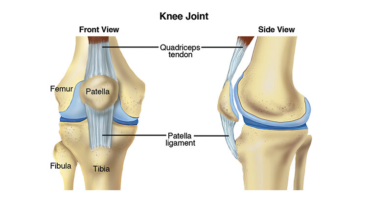
Osgood-Schlatter disease occurs when there is irritation to the top, front portion of the shin bone (tibia) where the tendon attached to the kneecap (patella) meets the shin bone. It occurs when there is an increased amount of stress placed upon the bones where the tendons attach. This is most often the result of increased activity levels by an adolescent athlete.

Our musculoskeletal system is made up of bones and surrounding soft tissue structures, including muscles, ligaments (which connect bone to bone), and tendons (which connect muscle to bone). These structures all play a role in helping us move.

During adolescence our bodies grow at a rapid rate. As our bodies develop, our bones are growing longer. Throughout this phase, our growth plates (epiphyseal plates) are susceptible to injury. A growth plate is the site at the end of a bone where new bone tissue is made and bone growth occurs. Females typically experience the most rapid growth between approximately 11 to 12 years of age, and males typically experience this growth surge between approximately 13 and 14 years of age. Males experience OS more frequently than females, likely due to an increased rate of sports participation.

Structures in our body might become irritated if they are asked to do more than they are capable of doing. Injuries can occur in an isolated event, but OS disease is most likely the cumulative effect of repeated trauma. OS is most frequently experienced in adolescents who regularly participate in running, jumping, and "cutting" (rapid changes in direction) activities.

When too much stress is present (ie, from rapid growth) and when the body is overworked (ie, either too much overall volume of exercise, or too much repetition), the top of the shin can become painful and swollen. As this condition progresses, the body’s response to bone stress can be an increase in bone production; an adolescent may begin to develop a boney growth that feels like a bump on the front of the upper shin.

OS can start as mild soreness, but can progress to long-lasting pain and limited function, if not addressed early and appropriately.

Back to Top

How Does it Feel?

With Osgood-Schlatter, you may experience:

  • Gradually worsening pain below your knee, at the top of the shin bone.

  • Pain that worsens with exercise.

  • Swelling and tenderness at the top of the shin.

  • A boney growth at the top of the shin.

  • Loss of strength in the quadriceps muscle (connecting the hip to the knee).

  • Increased tightness in the quadriceps muscle.

  • Loss of knee motion.

  • Discomfort with daily activities that use your knee, like kneeling, squatting, or walking up and down stairs.

Back to Top

How Is It Diagnosed?

Diagnosis of OS begins with a thorough medical history, including specific questions regarding athletic participation (sports played, frequency of practices/games, positions). Your physical therapist will assess different measures, such as sensation, motion, strength, flexibility, tenderness, and swelling. Your physical therapist will perform several tests specific to the knee joint, and may ask you to briefly demonstrate the activities or positions that cause your pain, such as walking, squatting, and stepping up or down stairs.

Because the knee and hip are both involved in these aggravating activities, your physical therapist will likely examine your hip as well. Other nearby areas, such as your feet and core, will also be examined to determine whether they, too, might be contributing to your knee condition.

If your physical therapist suspects there may be a more involved injury than increased stress-related irritation (ie, if there is a recent significant loss of motion or strength, or severe pain when the knee is moved), your therapist will likely recommend a referral to an orthopedic physician for diagnostic imaging, such as ultrasound, x-ray, or MRI.

Back to Top

How Can a Physical Therapist Help?

Once other conditions have been ruled out and OS is diagnosed, your physical therapist will work with you to develop an individualized treatment plan tailored to your specific knee condition and your goals. The goal of physical therapy is to accelerate your recovery and return to pain-free activity. There are many physical therapy treatments that have been shown to be effective in treating OS, and among them are:

Range of Motion Therapy. Your physical therapist will assess the motion of your knee and its surrounding structures, and design gentle exercises to help you work through any stiffness and swelling to return to a normal range of motion.

Strength Training. Your physical therapist will teach you exercises to strengthen the muscles around the knee so that each muscle is able to properly perform its job, and stresses are eased so the knee joint is properly protected.

Manual Therapy.Physical therapists are trained in manual (hands-on) therapy. If needed, your physical therapist will gently move your kneecap or patellar tendon and surrounding muscles as needed to improve their motion, flexibility, and strength. These techniques can target areas that are difficult to treat on your own. 

Pain Management.Your physical therapist may recommend therapeutic modalities, such as ice and heat, or a brace to aid in pain management.

Functional Training.Physical therapists are experts at training athletes to function at their best. Your physical therapist will assess your movements and teach you to adjust them to relieve any extra stress on the front of your knee.

Education. The first step to addressing your knee pain is rest. Your physical therapist will explain why this is important and develop a plan for your complete rehabilitation.

Back to Top

Can this Injury or Condition be Prevented?

Fortunately, there is much that can be done to prevent the cascade of events that lead to OS. Physical therapists focus on:

  • Educating coaches, parents, and athletes on guidelines for sports participation, explaining common causes of overuse injuries, and providing strategies for prevention.

  • Educating athletes on the risks of playing through pain.

  • Scheduling adequate rest time to recover between athletic events.

  • Tracking a young athlete’s growth curves (height, weight, BMI) to identify periods of increased injury risk.

  • Developing an athlete-specific flexibility and strengthening routine to be followed throughout the athletic season.

  • Encouraging consultation with a physical therapist whenever symptoms appear.

Back to Top

Real Life Experiences

Caleb is a 13-year-old boy who has been playing basketball since he was in the first grade. When younger, Caleb played only during the winter season. Over the course of the last year, however, he has attended 2 basketball camps during the summer, played on his middle-school team during the winter, and is now playing AAU basketball in the spring.

Caleb also chose to join the track team this spring, competing in the high jump and sprint events to improve his basketball skills. Over the past 3 months, Caleb has grown 2 inches, and both he and his basketball coaches are excited about his recent growth.

Recently, Caleb has been busy playing in weekly AAU tournaments with 1 to 2 track meets during the week. But when he got home from track practice on Monday, he told his dad that his leg was hurting. He said that it had begun getting sore while playing basketball over the weekend, but he didn’t want to tell his coach because he wanted to continue to play. Now he feels like the top of his shin is tender to touch, and he is unable to fully bend his knee without increased pain.

His dad realizes that this is more than the expected postactivity soreness; he immediately calls their local physical therapist.

Caleb's physical therapist takes his health history and performs an extensive examination. It becomes clear that Caleb has not scheduled appropriate rest times between his athletic activities, and that he is experiencing a growth spurt. The physical examination reveals that the top of Caleb’s shin is very tender, the area around his knee is swollen, and he has lost knee motion and strength. OS is diagnosed.

Together, Caleb and his physical therapist, father, and basketball and track coaches develop a treatment plan to help him return to pain-free sport participation. It begins with a 2-week period of rest where Caleb performs only minimal running exercise, and works regularly with his physical therapist on stretching, strengthening, balance, and coordination exercises, and on improving his squatting movements.

While at basketball practice, Caleb continues to work on his ball handling and free-throw shooting, activities that won't increase his knee pain.

After 2 weeks, when his knee is less tender, Caleb, his physical therapist, and his coaches develop a plan for his gradual return to full participation in track and basketball. They help Caleb understand how important it is to be honest about his knee pain, and to communicate with his coach if it starts to bother him again.

A month later, Caleb is back participating in all his track and basketball activities. He has changed his routine to allow for adequate warm-up time before and after each practice, and sufficient rest periods between activities. He makes sure his dad or coaches are keeping track of how much time he spends at practice and at rest.

Caleb decides to spend more time during the week working on his stretching, so he can reduce any risk of pain as he continues to grow. At the end of the season, Caleb’s AAU team wins the state championship—and he sets a new personal-best record in both the high jump and the 100-meter sprint!

Back to Top

What Kind of Physical Therapist Do I Need?

All physical therapists are prepared through education and clinical experience to treat a variety of conditions or injuries. You may want to consider:

  • A physical therapist who is experienced in treating people with orthopedic or musculoskeletal injuries.

  • A physical therapist who is a board-certified specialist or has completed a residency in orthopedic or sports physical therapy, as the therapist will have advanced knowledge, experience, and skills that apply to an athletic population.

You can find physical therapists that have these and other credentials by using Find a PT, the online tool built by the American Physical Therapy Association to help you search for physical therapists with specific clinical expertise in your geographic area.

General tips when you're looking for a physical therapist (or any other health care provider):

  • Get recommendations from family and friends or from other health care providers.

  • When you contact a physical therapy clinic for an appointment, ask about the physical therapists' experience in helping young athletes with knee pain.

  • During your first visit with the physical therapist, be prepared to describe your symptoms in as much detail as possible, and report activities that make your symptoms worse.

Back to Top

Further Reading

The American Physical Therapy Association (APTA) believes that consumers should have access to information that could help them make health care decisions, and also prepare them for a visit with their health care provider.

The following articles provide some of the best scientific evidence related to physical therapy treatment of athletic injuries. The articles report recent research and give an overview of the standards of practice both in the United States and internationally. The article titles are linked either to a PubMed* abstract of the article or to free full text, so that you can read it or print out a copy to bring with you to your health care provider.

Whitmore A. Osgood-Schlatter disease. JAAPA. 2013;26(10):51–52. Article Summary on PubMed.

Maffulli N, Longo UG, Spiezia F, Denaro V. Aetiology and prevention of injuries in elite young athletes. Med Sport Sci. 2011;56:187–200. Article Summary on PubMed.

Stein CJ, Micheli LJ. Overuse injuries in youth sports. Phys Sportsmed. 2010;38(2):102–108. Article Summary on PubMed.

* PubMed is a free online resource developed by the National Center for Biotechnology Information (NCBI). PubMed contains millions of citations to biomedical literature, including citations in the National Library of Medicine’s MEDLINE database.

 Authored by Allison Mumbleau, PT, DPT, SCS. Reviewed by the MoveForwardPT.com editorial board.


Patellofemoral Knee Pain

Patellofemoral pain syndrome (PFPS) refers to pain at the front of the knee, in and around the kneecap (patella). PFPS is one of the most common types of knee pain experienced in the United States, particularly among athletes, active teenagers, older adults, and people who perform physical labor. Patellofemoral pain affects more women than men and accounts for 20% to 25% of all reported knee pain. Physical therapists design exercise and treatment programs for people experiencing PFPS to help them reduce their pain, restore normal movement, and avoid future injury.

Current research indicates that PFPS is an "overuse syndrome," which means that it may result from repetitive or excessive use of the knee. Other contributing factors may include:

  • Weakness, tightness, or stiffness in the muscles around the knee and hip

  • An abnormality in the way the lower leg lines up with the hip, knee, and foot

  • Improper tracking of the kneecap

These conditions can interfere with the ability of the kneecap to glide smoothly on the femur (the bone that connects the knee to the thigh) in the femoral groove (situated along the thigh bone) during movement. The friction between the undersurface of the kneecap and the femur causes the pain and irritation commonly seen in PFPS. The kneecap also may fail to track properly in the femoral groove when the quadriceps muscle on the inside front of the thigh is weak, and the hip muscles on the outside of the thigh are tight. The kneecap gets pulled in the direction of the tight hip muscles and can track or tilt to the side, which irritates the tissues around the kneecap.

PFPS often occurs in people who are physically active or who have suddenly increased their level of activity, especially when that activity involves repeated knee motion, such as running, stair climbing, squatting, or repeated carrying of heavy loads. Older adults may experience age-related changes that cause the cartilage on the undersurface of the kneecap to wear out, resulting in pain and difficulty completing daily tasks without pain.

PatellofemoralPain_SM.jpg


How Does it Feel?

People with PFPS may experience:

  • Pain when walking up or down stairs or hills

  • Pain when walking on uneven surfaces

  • Pain that increases with activity and improves with rest

  • Pain that develops after sitting for long periods of time with the knee bent

  • A "crack" or "pop" when bending or straightening the knee

How Is It Diagnosed?

Your physical therapist will review your health history, perform a thorough examination, and conduct a series of tests to evaluate the knee. Your therapist may observe the alignment of your feet, analyze your walking and running patterns, and test the strength of your hip and thigh muscles to find out whether there is a weakness or imbalance that might be contributing to your pain. Your physical therapist also will check the flexibility of the muscles in your leg, paying close attention to those that attach at the knee.

Generally, X-rays are not needed to diagnose PFPS. Your physical therapist may consult with an orthopedic physician who may order an X-ray to rule out other conditions.

How Can a Physical Therapist Help?

After a comprehensive evaluation, your physical therapist will analyze the findings and, if PFPS is present, your therapist will prescribe an exercise and rehabilitation program just for you. Your program may include:

Strengthening exercises. Your physical therapist will teach you exercises targeted at the hip (specifically, the muscles of the buttock and thigh), the knee (specifically, the quadriceps muscle located on the front of your thigh that straightens your knee), and the ankle. Strengthening these muscles will help relieve pressure on the knee, as you perform your daily activities.

Stretching exercises. Your physical therapist also will choose exercises to gently stretch the muscles of the hip, knee, and ankle. Increasing the flexibility of these muscles will help reduce any abnormal forces on the knee and kneecap.

Positional training. Based on your activity level, your physical therapist may teach you proper form and positioning when performing activities, such as rising from a chair to a standing position, stair climbing, squatting, or lunging, to minimize excessive forces on the kneecap. This type of training is particularly effective for athletes.

Cross-training guidance. PFPS is often caused by overuse and repetitive activities. Athletes and active individuals can benefit from a physical therapist’s guidance about proper cross-training techniques to minimize stress on the knees.

Taping or bracing. Your physical therapist may choose to tape the kneecap to reduce your pain and retrain your muscles to work efficiently. There are many forms of knee taping, including some types of tape that help align the kneecap and some that just provide mild support to irritated tissues around it. In some cases, a brace may be required to hold the knee in the best position to ensure proper healing.

Electrical stimulation. Your physical therapist may prescribe treatments with gentle electrical stimulation to reduce pain and support the healing process.

Activity-based exercises. If you are having difficulty performing specific daily activities, or are an athlete who wants to return to a specific sport, your physical therapist will design individualized exercises to rebuild your strength and performance levels.

Fitting for an orthosis. If the alignment and position of your foot and arch appear to be contributing to your knee pain, your physical therapist may fit you with a special shoe insert called an orthosis. The orthosis can decrease the stress to your knee caused by low or high arches.

Can this Injury or Condition be Prevented?

PFPS is much easier to treat if it is caught early. Timely treatment by a physical therapist may help stop any underlying problems before they become worse. If you are experiencing knee pain, contact a physical therapist immediately. 

Your physical therapist can show you how to adjust your daily activities to safeguard your knees, and teach you exercises to do at home to strengthen your muscles and bones—and help prevent PFPS.

Physical therapists can assess athletic footwear and recommend proper choices for runners and daily walkers alike. Wearing the correct type of shoes for your activity and changing them when they are no longer supportive is essential to injury prevention.

Real Life Experiences

Amelia is a 25-year-old office assistant who loves to start her day with a 5-mile run. Over the past 6 months, she has been training for her first marathon. She began by training on very flat ground and has just moved to a hilly area.

Last week, Amelia began feeling pain in the front of her left knee when running downhill. Today, she had to stop running after 3 miles because of her knee pain. She called her physical therapist.

Amelia's physical therapist completes a comprehensive evaluation, including a screening for other possible conditions that might be causing her pain. He uses special tests to measure her strength and finds that she has weak hip muscles and tenderness around the kneecap. He determines that she has developed PFPS. Amelia is shocked to learn that she also has flat feet, and she’s not wearing the right supportive running shoes.

To begin her treatments, Amelia’s physical therapist applies special tape to the front of her knee to help reduce her pain, and instructs her in the use of ice to decrease her symptoms. He performs gentle movements of her kneecap and the surrounding tissues to help increase mobility and decrease pain. He teaches her special exercises to gently strengthen the weak muscles that support the knee.

He also designs a specific home-exercise program for Amelia to perform between sessions. He provides information about proper shoe choices for her foot and body type, and advises her to purchase shoes that will give her feet the right type of support. He also recommends that she try deep-water running or swimming for a week instead of her regular running program, until her condition improves.

After her first week of physical therapy, Amelia notices a decrease in her pain and an increased ability to walk up and down stairs without pain. Her physical therapist approves her new footwear, and adds more challenging exercises to her session and her home program. He gives her the go-ahead to race-walk. She applies ice only when she has pain.

After 2 weeks, Amelia reports she is feeling even less pain. Her physical therapist continues to increase the intensity of her exercises, and she starts to run again—but only on flat surfaces and short distances combined with longer walk intervals.

After a few more weeks of therapy, Amelia occasionally feels only slight twinges of pain and gradually resumes her prior level of training. Her physical therapist recommends continuation of her stretching and strengthening exercises, and discharges her from physical therapy.

A few months later, Amelia completes her first marathon pain free. She is thrilled to learn that her time was a personal best!

This story was based on a real-life case. Your case may be different. Your physical therapist will tailor a treatment program to your specific case.

What Kind of Physical Therapist Do I Need?

All physical therapists are prepared through education and experience to treat a variety of conditions or injuries. You may want to consider:

  • A physical therapist who is experienced in treating people with orthopedic, or musculoskeletal, problems.

  • A physical therapist who is a board-certified clinical specialist or who has completed a residency or fellowship in orthopedic physical therapy and has advanced knowledge, experience, and skills that may apply to your condition.

You can find physical therapists who have these and other credentials by using Find a PT, the online tool built by the American Physical Therapy Association to help you search for physical therapists with specific clinical expertise in your geographic area.

General tips when you're looking for a physical therapist:

  • Get recommendations from family and friends or from other health care providers.

  • When you contact a physical therapy clinic for an appointment, ask about the physical therapist's experience in helping people with patellofemoral pain syndrome.

During your first visit with the physical therapist, be prepared to describe your symptoms in as much detail as possible, and say what makes your symptoms worse.

Further Reading

The American Physical Therapy Association (APTA) believes that consumers should have access to information that could help them make health care decisions and also prepare them for their visit with their health care provider.

The following articles provide some of the best scientific evidence related to physical therapy treatment of patellofemoral pain syndrome. The articles report recent research and give an overview of the standards of practice both in the United States and internationally. The article titles are linked either to a PubMed* abstract of the article or to free access of the full article, so that you can read it or print out a copy to bring with you to your health care provider.

Tevhen DS, Robertson J. Knee pain: strengthen my hips? But it's my knees that hurt! J Orthop Sports Phys Ther. 2011-41-571. Article Summary on PubMed.

Davis IS, Powers CM. Patellofemoral pain syndrome: proximal, distal and local factors, an international retreat, April 30-May 2, 2009, Fells Point, Baltimore, MD. J Orthop Sports Phys Ther. 2010;40:A1–A16. Article Summary on PubMed.

Fukuda TY, Rossetto FM, Magalhaes E, et al. Short-term effects of hip abductors and lateral rotators strengthening in females with patellofemoral pain syndrome: a randomized controlled clinical trial. J Orthop Sports Phys Ther. 2010;40:736–742. Article Summary on PubMed.

Dixit S, DiFiori JP, Burton M, Mines B. Management of patellofemoral pain syndrome. Am Fam Physician. 2007;75:194–202. Free Article.

Powers CM, Ward SR, Chan LD, et al. The effect of bracing on patella alignment and patellofemoral joint contact area. Med Sci Sports Exerc. 2004;36:1226-1232. Article Summary on PubMed.

Bizzini M, Childs JD, Piva SR, Delitto A. Systematic review of the quality of randomized controlled trials for patellofemoral pain syndrome. J Ortho Sports Phys Ther. 2003;33:4–20. Article Summary on PubMed.

Crossley K, Bennell K, Green S, et al. Physical therapy for patellofemoral pain: a randomized, double-blinded, placebo-controlled trial. Am J Sports Med. 2002;30:857–865. Article Summary on PubMed.


* PubMed is a free online resource developed by the National Center for Biotechnology Information (NCBI).  PubMed contains millions of citations to biomedical literature, including citations in the National Library of Medicine’s MEDLINE database.

 Authored by Christopher Bise, PT, MS, DPT. Revised by Julie Mulcahy, PT. Reviewed by the MoveForwardPT.com editorial board.


Total Knee Replacement (Arthroplasty)

The knee is the most commonly replaced joint in the body. The decision to have knee replacement surgery is one that you should make in consultation with your orthopedic surgeon and your physical therapist. Usually, total knee replacement surgery is performed when people have:

  • Knee joint damage due to osteoarthritisrheumatoid arthritis, other bone diseases, or fracture that has not responded to more conservative treatment options

  • Knee pain or alignment problems in the leg that cause difficulty with walking or performing daily activities, which have not responded to more conservative treatment options

What is a Total Knee Replacement (TKR)?

A total knee replacement (TKR), also known as total knee arthroplasty, involves removing the arthritic parts of the bones at the knee joint (the tibia, sometimes called the shin bone; the femur, or thigh bone; and the patella, or kneecap) and replacing them with artificial parts. These parts consist of a metal cap at the end of the femur and a cemented piece of metal in the tibia with a plastic cap on it to allow the surfaces to move smoothly. When appropriate, the back part of the kneecap also may be replaced with a smooth plastic surface.

KneeReplacement-SM.jpg

How Can a Physical Therapist Help?

The physical therapist is an integral part of the team of health care professionals who help people receiving a total knee replacement regain movement and function, and return to daily activities. Your physical therapist can help you prepare for and recover from surgery, and develop an individualized treatment program to get you moving again in the safest and most effective way possible.

Before Surgery

The better physical shape you are in before TKR surgery, the better your results will be (especially in the short term). A recent study has shown that even 1 visit with a physical therapist prior to surgery can help reduce the need for short-term care after surgery, such as a short stay at a skilled nursing facility, or a home health physical therapy program.

Before surgery, your physical therapist may:

  • Teach you exercises to improve the strength and flexibility of the knee joint and surrounding muscles.

  • Demonstrate how you will walk with assistance after your operation, and prepare you for the use of an assistive device, such as a walker.

  • Discuss precautions and home adaptations with you, such as removing loose accent rugs that could cause you to “catch” your leg on them when maneuvering with an assistive device, or strategically placing a chair so that you can sit instead of squatting to get something out of a low cabinet. It is always easier to make these modifications before you have TKR surgery.

Longer-term adjustments that are recommended prior to surgery include:

  • Stopping smoking. Seek assistance or advice from your physician on stopping smoking, as you schedule and plan for your surgery. Being tobacco-free will improve your healing process following surgery.

  • Losing weight. Losing excess body weight may help you recover more quickly, and help improve your function and overall results following surgery.

Immediately Following Surgery

You may stay in the hospital for a few days following surgery, or you may even go home on the same day, depending on your condition. If you have other medical conditions, such as diabetes or heart disease, you might need to stay in the hospital or go to a skilled nursing facility for a few days before returning home. While you are in the hospital, a physical therapist will:

  • Educate you on applying ice, elevating your leg, and using compression wraps or stockings to control swelling in the knee area and help the incision heal.

  • Teach you breathing exercises to help you relax, and show you how to safely get in and out of bed and a chair.

  • Show you how to walk with a walker or crutches, and get in and out of a car.

  • Help you continue to do the flexibility and strengthening exercises that you learned before your surgery.

As You Begin to Recover

The goal of the first 2 weeks of recovery is to manage pain, decrease swelling, heal the incision, restore normal walking, and initiate exercise. Following those 2 weeks, your physical therapist will tailor your range-of-motion exercises, progressive muscle-strengthening exercises, body awareness and balance training, functional training, and activity-specific training to address your specific goals and get you back to the activities you love!

Range-of-motion exercises. Swelling and pain can make you move your knee less. Your physical therapist can teach you safe and effective exercises to restore movement (range of motion) to your knee, so that you can perform your daily activities.

Strengthening exercises. Weakness of the muscles of the thigh and lower leg could make you need to still use a cane when walking, even after you no longer need a walker or crutches. Your physical therapist can determine which strengthening exercises are right for you.

Body awareness and balance training. Specialized training exercises help your muscles "learn" to respond to changes in your world, such as uneven sidewalks or rocky ground. When you are able to put your full weight on your knee without pain, your physical therapist may add agility exercises (such as turning and changing direction when walking, or making quick stops and starts) and activities using a balance board that challenge your balance and knee control. Your program will be based on the physical therapist’s examination of your knee, on your goals, and on your activity level and general health.

Functional training. When you can walk freely without pain, your physical therapist may begin to add activities that you were doing before your knee pain started to limit you. These might include community-based actions, such as crossing a busy street or getting on and off an escalator. Your program will be based on the physical therapist's examination of your knee, on your goals, and on your activity level and general health.

The timeline for returning to leisure or sports activities varies from person-to-person; your physical therapist will be able to estimate your unique timeline based on your specific condition.

Activity-specific training. Depending on the requirements of your job or the type of sports you play, you might need additional rehabilitation that is tailored to your job activities (such as climbing a ladder) or sport activities (such as swinging a golf club) and the demands that they place on your knee. Your physical therapist can develop an individualized rehabilitation program for you that takes all of these demands into account.

Can this Injury or Condition be Prevented?

If you have knee pain, you may be able to delay the need for surgery by working with a physical therapist to improve the strength and flexibility of the muscles that support and move the knee. This training could even help you avoid surgery altogether. Participating in an exercise program designed by a physical therapist can be one of your best protections against knee injury. And staying physically active in moderately intense physical activities and controlling your weight through proper diet might help reduce the risk of osteoarthritis of the knee getting worse.

Real Life Experiences

Carmella is a 67-year-old grandmother of 3 who has had osteoarthritis in her right knee for a few years. She used to take care of her grandchildren after school each day before her daughter got home from work. Then Carmella's knee became so painful that she could no longer walk up and down stairs or stand for long periods of time. She also had a lot of difficulty getting up from a chair. She had to tell her daughter that she couldn't take care of her grandchildren anymore. She decided to see a physical therapist.

Carmella’s physical therapist began her first session by asking detailed questions about her knee, such as what other treatments Carmella had tried and the outcomes of those treatments. Carmella said she had seen an orthopedic surgeon who had suggested injections, which helped reduce her pain for a period of time. Her physical therapist then asked her how her current knee pain affected her ability to do the things she wanted to do. Carmella said it made her unable to care for her grandchildren, participate in a regular walking program for fitness, or do the things she enjoyed for recreation.

Her physical therapist then took some measurements of her knee range of motion and strength and conducted tests to get a better idea of what was generating her pain. He suggested that she consult with an orthopedic surgeon. After carefully reviewing her condition and learning about her previous treatments and current activity limitations, the surgeon suggested it was time for a total knee replacement. Carmella agreed. The surgeon scheduled the procedure for 1 month later.

To prepare for surgery, Carmella’s physical therapist taught her strengthening and stretching exercises, showed her how to use crutches following surgery, and advised her on preparing her home environment to make it safe post surgery.

The first day after her surgery, a hospital-based physical therapist came to Carmella's room to begin a gentle recovery program. She showed Carmella how to bend and straighten her knee and how to tense and then relax and release her knee, calf, and hip muscles to strengthen them. She then helped Carmella practice sitting at the edge of her hospital bed and standing up using crutches.

The second day after surgery, Carmella started walking with crutches with the physical therapist’s assistance, putting a little weight on her right leg. The physical therapist also instructed her in some gentle leg-strengthening exercises.

On the third day after surgery, Carmella was able to walk using her crutches, monitored by the physical therapist but without her help, in the hospital hallways and up and down a few stairs. Her physical therapist designed an at-home exercise program just for her, and taught it to her. Carmella was discharged home with a pair of crutches.

Once Carmella returned home, a home-care physical therapist regularly visited her at her house to continue her rehabilitation. As she improved, he prescribed more challenging exercises for her that added weights for strengthening. Carmella also began to practice walking with a cane instead of her crutches.

Two weeks after her surgery, Carmella began going to outpatient physical therapy. Her pain progressively decreased and she had noticeable improvements in her knee range of motion and the strength of her lower body. She and her physical therapist developed a plan that would help allow her to get back to her recreational activities as well as allow her to care for her grandchildren.

A few weeks laterCarmella felt hardly any pain in her knee. She could walk without using a cane, but still needed to use a handrail when going up or down stairs. At times, her knee felt "shaky." She told her physical therapist she was still not comfortable taking care of her grandchildren because of these remaining challenges.

Carmella's physical therapist instructed her in more aggressive strengthening and movement exercises for her hips, knees, and ankles. She also worked with her on improving her stair climbing, balance, and agility. Carmella began to feel more confident walking up and down stairs, getting in and out of her car and driving, and performing other daily activities. She felt that her new knee was much more stable.

A few weeks later, Carmella was able to take care of her grandchildren again! She also joined a health club that offered exercise programs for older adults, so she could maintain the benefits she had gained from her physical therapy.

This story was based on a real-life case. Your case may be different. Your physical therapist will tailor a treatment program to your specific case.

What Kind of Physical Therapist Do I Need?

Although all physical therapists are prepared through education and experience to treat people who have a TKR, you may want to consider:

  • A physical therapist who is experienced in treating people with orthopedic, or musculoskeletal, problems

  • A physical therapist who is a board-certified clinical specialist or who has completed a residency or fellowship in orthopedic physical therapy, giving the physical therapist advanced knowledge, experience, and skills that may apply to your condition

You can find physical therapists who have these and other credentials by using Find a PT, the online tool built by the American Physical Therapy Association to help you search for physical therapists with specific clinical expertise in your geographic area.

General tips when you're looking for a physical therapist:

  • Get recommendations from family and friends or from other health care providers.

  • When you contact a physical therapy clinic for an appointment, ask about the physical therapist's experience in helping people with TKR.

During your first visit with the physical therapist, be prepared to describe your symptoms in as much detail as possible, and say what makes your symptoms worse.

Further Reading

The American Physical Therapy Association (APTA) believes that consumers should have access to information that could help them make health care decisions and also prepare them for their visit with their health care provider.

The following articles provide some of the best scientific evidence about physical therapist treatment of TKR. The articles report recent research and give an overview of the standards of practice for treatment both in the United States and internationally. The article titles are linked either to a PubMed abstract (summary) of the article or to free access of the entire article, so that you can read it or print out a copy to bring with you when you see your health care provider.

Harmelink KE, Zeegers AV, Hullegie W, et al. Are there prognostic factors for one-year outcome after total knee arthroplasty: a systematic review. J Arthroplasty. 2017 August 1 [Epub ahead of print]. doi: 10.1016/j.arth.2017.07.011. Article Summary in PubMed.

Pua YH, Seah FJ, Poon CL, et al. Age- and sex-based recovery curves to track functional outcomes in older adults with total knee arthroplasty. Age Ageing. 2017 August 30 [Epub ahead of print]. doi: 10.1093/ageing/afx148. Article Summary in PubMed.

Sobh AH, Siljander MP, Mells AJ, et al. Cost analysis, complications, and discharge disposition associated with simultaneous vs staged bilateral total knee arthroplasty. J Arthroplasty. 2017 September 13 [Epub ahead of print]. doi: 10.1016/j.arth.2017.09.004. Article Summary in PubMed.

Bistolfi A, Zanovello J, Ferracini R, et al. Evaluation of the effectiveness of neuromuscular electrical stimulation after total knee arthroplasty: a meta-analysis. Am J Phys Med Rehabil. 2017 October 7 [Epub ahead of print]. Article Summary in PubMed.

Otero-López A, Beaton-Comulada D. Clinical considerations for the use lower extremity arthroplasty in the elderly. Phys Med Rehabil Clin N Am. 2017;28(4):795–810. Article Summary in PubMed.

Loyd BJ, Jennings JM, Judd DL, et al. Influence of hip abductor strength on functional outcomes before and after total knee arthroplasty: post hoc analysis of a randomized controlled trial. Phys Ther. 2017;97(9):896–903. Article Summary in PubMed.

Piva SR, Teixeira PE, Almeida GJ, et al. Contribution of hip abductor strength to physical function in patients with total knee arthroplasty. Phys Ther. 2011;91:225–233. Free Article.

Dowsey MM, Liew D, Choong PF. The economic burden of obesity in primary total knee arthroplasty. Arthritis Care Res(Hoboken). 2011;63(10):1375–1381. Article Summary on PubMed.

Piva SR, Gil AB, Almeida GJ, et al. A balance exercise program appears to improve function for patients with total knee arthroplasty: a randomized clinical trial. Phys Ther. 2010;90:880–894. Free Article.

Bade MJ, Kohrt WM, Stevens-Lapsley JE. Outcomes before and after total knee arthroplasty compared to healthy adults. J Ortho Sports Phys Ther. 2010;40:559–567. Free Article.

Walls RJ, McHugh G, O’Gorman DJ, et al. Effects of preoperative neuromuscular electrical stimulation on quadriceps strength and functional recovery in total knee arthroplasty: a pilot study. BMC Musculoskelet Disord. 2010;11:119. Free Article.

Topp R, Swank AM, Quesada PM, et al. The effect of prehabilitation exercise on strength and functioning after total knee arthroplasty. PM R. 2009;1:729–735. Article Summary on PubMed.

Kirkley A, Birmingham TB, Litchfield RB, et al. A randomized trial of arthroscopic surgery for osteoarthritis of the knee [published correction appears in: N Engl J Med. 2009;361:2004]. N Engl J Med. 2008;359:1097–1107. Free Article.

Minns Lowe CJ, Barker KL, Dewey M, Sackley CM. Effectiveness of physiotherapy exercise after knee arthroplasty for osteoarthritis: systematic review and meta-analysis of randomised controlled trials. BMJ. 2007;335:812. Free Article.

Moffet H, Collet JP, Shapiro SH, et al. Effectiveness of intensive rehabilitation on functional ability and quality of life after first total knee arthroplasty: a single-blind randomized controlled trial. Arch Phys Med Rehabil. 2004;85:546–556. Free Article.

Deyle GD, Henderson NE, Matekel RL, et al. Effectiveness of manual physical therapy and exercise in osteoarthritis of the knee: a randomized, controlled trial. Ann Intern Med. 2000;132:173–181. Free Article.

 *PubMed is a free online resource developed by the National Center for Biotechnology Information (NCBI). PubMed contains millions of citations to biomedical literature, including citations in the National Library of Medicine’s MEDLINE database.

Authored by Anne Reicherter, PT, DPT, PhDThe author is a board-certified clinical specialist in orthopaedic physical therapyReviewed by the MoveForwardPT.com editorial board.


Medial Collateral Ligament (MCL) Injury

The medial collateral ligament (MCL) is the most commonly damaged ligament in the knee. The MCL can be sprained or torn as a result of a blow to the outer side of the knee, by twisting the knee, or by quickly changing directions while walking or running. MCL injury most often occurs in athletes, although nonathletes can also be affected. A physical therapist treats an MCL sprain or tear to reduce pain, swelling, stiffness, and any associated weakness in the knee or lower extremity.

What is an MCL Injury?

The MCL is a small, thick band of tissue on the inner side of the knee joint. It connects two bones—the thighbone and the shin bone—preventing the knee from bending inward toward the other knee. When the knee is hit on the outer side of the leg (eg, the left side of the left leg), or if the knee is twisted violently, the MCL can overstretch resulting in a partial or complete tear. MCL injuries commonly occur in football players who get "clipped" or hit on the outer side of the knee. Other causes may include twisting and turning while skiing, blows received on the soccer field, trauma experienced in a car accident, or simply turning the knee sharply while the foot is planted on the ground. Healing times vary from a couple of weeks to a couple of months, depending on the severity of the injury.

How Does it Feel?

When you experience an MCL injury, you may feel:

  • Pain on the inner side of the knee

  • Swelling and bruising at the inner side of the knee

  • Swelling that spreads to the rest of the knee joint in 1 or 2 days following injury

  • Stiffness in the knee

  • Difficulty or pain when trying to bend or straighten the knee

  • An unstable feeling, as though the knee may give out or buckle

  • Pain or difficulty walking, sitting down, rising from a chair, or climbing stairs

Signs and Symptoms

With an MCL injury, you may experience

  • A "popping" sound as the injury occurs

  • Pain and swelling in your knee

  • Difficulty moving your knee

  • Difficulty bearing weight on your leg for walking or getting up from a chair

How Is It Diagnosed?

If you see your physical therapist first, the therapist will conduct a thorough evaluation that includes taking your health history. Your therapist will also ask you detailed questions about your injury, such as:

  • Did you feel pain or hear a "pop" when you injured your leg?

  • Did you turn your leg with your foot planted on the ground?

  • Did you change direction quickly while running?

  • Did you receive a direct hit to the leg while your foot was planted on the ground?

  • Did you see swelling around the knee in the first 2 to 3 hours following the injury?

  • Does your knee feel like buckling or giving way when you try to use it?

Your physical therapist also will perform special tests to help determine the likelihood that you have an MCL injury. Your therapist will gently press on the outside of your knee while it is slightly bent as well as when it is fully straight to test the strength of the ligament. The therapist will also check the inner side of your knee for tenderness and swelling and measure for swelling with a tape measure. The therapist may use additional tests to determine if other parts of your knee are injured, and will also observe how you are walking.

To provide a definitive diagnosis, your therapist may collaborate with an orthopedic physician or other health care provider. The orthopedic physician may order further tests, such as magnetic resonance imaging (MRI), to confirm the diagnosis and to rule out other damage to the knee. It also helps to determine whether surgery is required. MRI is not required in all cases but may be ordered. Your therapist or doctor may recommend a knee brace, a knee immobilizer, or crutches to reduce pain if the MCL injury is severe.

How Can a Physical Therapist Help?

Your physical therapist will work with you to design a specific treatment program that will speed your recovery, including exercises and treatments you can do at home. Physical therapy will help you return to your normal lifestyle and activities.

The First 24-48 Hours

Your physical therapist may advise you to:

  • Rest the area by avoiding walking or any activity that causes pain. Crutches and a knee brace may be recommended to reduce further strain on the MCL when walking.

  • Apply ice packs to the area for 15-20 minutes every 2 hours.

  • Compress the area with an elastic bandage wrap.

  • Consult with a physician for further services such as medication or diagnostic tests.


Reduce Pain

Your physical therapist may use different types of treatments and technologies to control and reduce your pain, including ice, heat, ultrasound, electrical stimulation, taping, exercises, and hands-on therapy such as massage.

Improve Motion

Your physical therapist will choose specific activities and treatments to help restore normal movement in the knee and leg. These might begin with passive motions that the therapist performs for you to gently move your leg and knee joint, and progress to active exercises and stretches that you do yourself.

Improve Strength

Certain exercises will aid healing at each stage of recovery; your physical therapist will choose and teach you the correct exercises and equipment to steadily restore your strength and agility. These may include using cuff weights, stretchy bands, weight-lifting equipment, and cardio-exercise equipment such as treadmills or stationary bicycles.

Improve Balance

Regaining your sense of balance is important after an injury. Your physical therapist will teach you exercises to improve your balance skills.

Speed Recovery Time

Normal healing of time is a few weeks to a few months, depending on which tissues are injured and how severely they are injured. Your physical therapist is trained and experienced in choosing the right treatments and exercises to help you heal, return to your normal lifestyle, and reach your goals faster than you are likely to do on your own.

Return to Activities

Your physical therapist will discuss your goals with you and use them to set your work, sport, and homelife recovery goals. The therapist will design your treatment program to help you reach those goals in the safest, fastest, and most effective way possible. Your physical therapist will apply hands-on therapy, such as massage, and teach you exercises, work retraining activities, and sport-specific techniques and drills to help you achieve your goals.

Prevent Future Injury

Your physical therapist can recommend a home exercise program to strengthen and stretch the muscles around your knee, upper leg, and abdomen to help prevent future injury. These may include strength and flexibility exercises for the leg, knee, and core muscles.

If Surgery Is Necessary

Surgery is rarely necessary in the case of an MCL injury. If surgery is needed, you will follow a recovery program over several weeks guided by your physical therapist, who will help you minimize pain, regain motion, strength, and return to normal activities as quickly as possible after surgery.

Can this Injury or Condition be Prevented?

To help prevent a recurrence of the injury, your physical therapist may advise you to:

  • Learn how to not let your knees collapse in toward each other when jumping, running, or turning quickly

  • Practice balance and agility exercises and drills

  • Always warm up before starting a sport or heavy physical activity

  • Follow a consistent strength and flexibility exercise program to maintain good physical conditioning, even in a sport's off-season

  • Wear shoes that are in good condition and fit well

Real Life Experiences

Mark is a 35-year-old accountant who is an avid bowler on the weekends. He lives with his 100-lb Rottweiler dog. One morning, as Mark was quickly turning a corner into the kitchen to grab a ringing phone, his dog ran the other way and accidentally hit Mark’s knee on the outer side of his right leg. Mark lost his balance and fell sideways. His right foot got caught underneath the dog as his body fell to the right, forcing the outer side of the knee to buckle and the inner side of the knee to overstretch. Mark felt a sharp pain on the inner side of his knee, and fell to the ground. Mark felt immediate tenderness on the inner side of his knee, and he could not straighten or bend it.

Mark was able to see his physical therapist that day. The physical therapist performed special tests on the ligaments and cartilage in the knee. She found that just the MCL was injured, and that it was a mild sprain. She immediately applied ice and electrical stimulation to the area for 20 minutes. She wrapped Mark’s knee with a compressive wrap and instructed him to keep it elevated when he was sitting or lying down. She gave Mark crutches and taught him how to use them.

When Mark returned for his next visit, the physical therapist began gently moving the knee to reduce the stiffness. She taught Mark some exercises he could do at home to start improving his muscle strength. She helped him use equipment in the clinic to gently move, stretch, and strengthen his knee and leg.

Mark received physical therapy treatments for 2 weeks, after which he was able to walk and climb stairs with only a little discomfort. His therapist taught him a variety of balance and endurance exercises. By the third week, he was able to return to bowling, and walk around sharp corners in his house, while keeping a watchful eye on his energetic dog!

This story was based on a real-life case. Your case may be different. Your physical therapist will tailor a treatment program to your specific case.

What Kind of Physical Therapist Do I Need?

All physical therapists are prepared through education and experience to treat MCL injury. However, you may want to consider:

  • A physical therapist who is experienced in treating people with orthopedic injuries. Some physical therapists have a practice with an orthopedic focus.

  • A physical therapist who is a board-certified clinical specialist or who has completed a residency or fellowship in orthopedic or sports physical therapy. This therapist has advanced knowledge, experience, and skills that may apply to your condition.

You can find physical therapists that have these and other credentials by using Find a PT, the online tool developed by the American Physical Therapy Association to help you search for physical therapists with specific clinical expertise in your geographic area.

General tips when you're looking for a physical therapist (or any other health care provider):

  • Get recommendations from family and friends or from other health care providers.

  • When you contact a physical therapy clinic for an appointment, ask about the physical therapists' experience in helping people who have your type of injury.

  • During your first visit with the physical therapist, be prepared to describe your symptoms in as much detail as possible, and say what makes your symptoms worse.

Further Reading

The American Physical Therapy Association (APTA) believes that consumers should have access to information that could help them make health care decisions and also prepare them for their visit with their health care provider.

The following articles provide some of the best scientific evidence related to physical therapy treatment of MCL injury. The articles report recent research and give an overview of the standards of practice both in the United States and internationally. The article titles are linked either to a PubMed* abstract of the article or to free full text, so that you can read it or print out a copy to bring with you to your health care provider.

Frommer C, Masaracchio M. The use of patellar taping in the treatment of a patient with a medial collateral ligament sprain. N Am J Sports Phys Ther. 2009;4(2):60-69. Free Article.

Hunt SE, Herrera C, Cicerale S, et al. Rehabilitation of an elite olympic class sailor with MCL injury. N Am J Sports Phys Ther. 2009;4(3):123-131. Free Article.

Edson CJ. Conservative and postoperative rehabilitation of isolated and combined injuries of the medial collateral ligament.Sports Med Arthrosc. 2006;14(2):105-110. Article Summary on PubMed.

Azar FM. Evaluation and treatment of chronic medial collateral ligament injuries of the knee. Sports Med Arthrosc. 2006;14(2):84-90. Article Summary on PubMed.

Fung DT, Ng GY, Leung MC, Tay DK. Effects of a therapeutic laser on the ultrastructural morphology of repairing medial collateral ligament in a rat model. Lasers Surg Med. 2003;32(4):286-293. Article Summary on PubMed.

Reider B. Medial collateral ligament injuries in athletes. Sports Med. 1996;21(2):147-156. Article Summary on PubMed.

Paletta GA, Warren RF. Knee injuries and Alpine skiing: treatment and rehabilitation. Sports Med. 1994;17(6):411-423. ArticleSummary on PubMed.

*PubMed is a free online resource developed by the National Center for Biotechnology Information (NCBI).  PubMed contains millions of citations to biomedical literature, including citations in the National Library of Medicine’s MEDLINE database.

 Authored by Andrea Avruskin, PT, DPT. Reviewed by the MoveForwardPT.com editorial board.

Physical Therapist's Guide to Iliotibial Band Syndrome (ITBS or "IT Band Syndrome")

IliotibialBand_Small.jpg

Iliotibial band syndrome (ITBS) is one of the most common causes of knee pain, particularly in individuals involved in endurance sports. It accounts for up to 12% of running injuries and up to 24% of cycling injuries. ITBS is typically managed conservatively through physical therapy and temporary activity modification.

What is Iliotibial Band Syndrome (ITBS)?

Iliotibial band syndrome (ITBS) occurs when excessive irritation causes pain at the outside (or lateral) part of the knee. The iliotibial band (ITB), often referred to as the "IT band" is a type of soft tissue that runs along the side of the thigh from the pelvis to the knee. As it approaches the knee, its shape thickens as it crosses a prominent area of the thigh (femur) bone, called the lateral femoral condyle. Near the pelvis, it attaches to 2 important hip muscles, the tensor fascia latae (TFL) and the gluteus maximus.

Irritation and inflammation arise from friction between the ITB and underlying structures when an individual moves through repetitive straightening (extension) and bending (flexion) of the knee. Typically, ITBS pain occurs with overuse during activities such as running and cycling.

ITBS involves many lower extremity structures, including muscles, bones, and other soft tissues. Usually discomfort arises from:

  • Abnormal contact between the ITB and thigh (femur) bone
  • Poor alignment and/or muscular control of the lower body
  • Prolonged pinching (compression) or rubbing (shearing) forces during repetitive activities

The common structures involved in ITBS are:

  • Iliotibial band
  • Bursa (fluid-filled sack that sits between bones and soft tissues to limit friction)
  • Hip muscles

ITBS can occur in:

  • Athletes performing repetitive activities, such as squatting, and endurance sports such as running and cycling
  • Individuals who spend long periods of time in prolonged positions, such as sitting or standing for a long workday, climbing or squatting, or kneeling
  • Individuals who quickly start a new exercise regimen without proper warm-up or preparation

Back to Top


Signs and Symptoms

With ITBS, you may experience:

  • Stabbing or stinging pain along the outside of the knee
  • A feeling of the ITB “snapping” over the knee as it bends and straightens
  • Swelling near the outside of your knee
  • Occasionally, tightness and pain at the outside of the hip
  • Continuous pain following activity, particularly with walking, climbing, or descending stairs, or moving from a sitting to standing position

Pain is usually most intense when the knee is in a slightly bent position, either right before or right after the foot strikes the ground. This is the point where the ITB rubs the most over the femur.

Back to Top


How Is It Diagnosed?

Your physical therapist will ask you questions about your medical history and activity regimen. A physical examination will be performed so that your physical therapist can collect movement (range of motion), strength, and flexibility measurements at the hip, knee, and ankle.

When dealing with ITBS, it is also common for a physical therapist to use special tests and complete a movement analysis, which will provide information on the way that you move and how it might contribute to your injury. This could include assessment of walking/running mechanics, foot structure, and balance. Your therapist may have you repeat the activity that causes your pain to see firsthand how your body moves when you feel pain. If you are an athlete, your therapist might also ask you about your chosen sport, shoes, training routes, and exercise routine.

Typically, medical imaging tests, such as x-ray and MRI, are not needed to diagnosis ITBS.

Back to Top


How Can a Physical Therapist Help?

Your physical therapist will use treatment strategies to focus on:

Range of motion

Often, abnormal motion of the hip and knee and foot joint can cause ITBS because of how the band attaches to hip muscles. Your therapist will assess the motion of your injury leg compared with expected normal motion and the motion of the hip on your uninvolved leg.

Muscle strength

Hip and core weakness can contribute to ITBS. The "core" refers to the muscles of the abdomen, low back, and pelvis. Core strength is important, as a strong midsection will allow greater stability through the body as the arms and legs go through various motions. For athletes performing endurance sports, it is important to have a strong core to stabilize the hip and knee joints during repetitive leg motions. Your physical therapist will be able to determine which muscles are weak and provide specific exercises to target these areas.

Manual therapy

Many physical therapists are trained in manual therapy, which means they use their hands to move and manipulate muscles and joints to improve motion and strength. These techniques can target areas that are difficult to treat on your own.

Functional training

Even when an individual has normal motion and strength, it is important to teach the body how to perform controlled and coordinated movements so there is no longer excessive stress at the previously injured structures. Your physical therapist will develop a functional training program specific to your desired activity. This means creating exercises that will replicate your activities and challenge your body to learn the correct way to move.

Your physical therapist will also work with you to develop an individualized treatment program specific to your personal goals. He or she will offer tips to help you prevent your injury from reoccurring.

Back to Top


Can this Injury or Condition be Prevented?

Maintaining core and lower extremity strength and flexibility and monitoring your activity best prevents ITBS. It is important to modify your activity and contact your physical therapist soon after first feeling pain. Research indicates that when soft tissues are irritated and the offending activity is continued, the body does not have time to repair the injured area. This often leads to persistent pain, and the condition becomes more difficult to resolve.

Once you are involved in a rehabilitation program, your physical therapist will help you determine when you are ready to progress back to your previous activity level. He or she will make sure that your body is ready to handle the demands of your activities so that your injury does not return. You will also receive a program to perform at home that will help you maintain the improvements that you gained during rehabilitation.

Back to Top


Real Life Experiences

Sarah is a 31-year-old mother training for her first triathlon. With a young child at home, she has to squeeze in her training sessions early in the morning. She rarely has time to cool down or stretch after riding her bike or running because she has to get home before her child wakes up.

Sarah signs up for her first race and begins to increase her cycling and running in preparation. One day during the middle of a long run, she feels a sharp pain at the outside of her knee. It starts hurting with every step, and doesn't go away, even after she stops and stretches. Far from home, she has to finish her run despite the nagging pain. When she gets home, she puts ice on it, but for the rest of the day she has trouble going up and down stairs, or squatting to pick up her son, and feels pain when standing up after driving the car. The next day, she tries to ride her bike, but the knee pain is still there and feels worse.

Wisely, Sarah stops running and cycling and contacts her physical therapist.

Sarah's physical therapist conducts a comprehensive evaluation of her hip and knee motion, strength, balance, and running mechanics. She uses special tests and measures to determine if Sarah’s pain is related to her iliotibial band or if there are other problems occurring simultaneously. She talks with Sarah about her training routine, including equipment (shoes, position on the bike, etc), the routes she runs and their surfaces, and her stretching program. The therapist diagnoses Sarah with iliotibial band syndrome. She guides Sarah through specific exercises in the clinic, including manual stretching of the hip joint by the therapist, sidelying leg raises for hip strengthening, and single leg squats to promote integrated core, hip, knee, and ankle function. Sarah will also perform these exercises at home as a part of her daily exercise routine to maximize improvement and help ensure her sustainable success.

Sarah's physical therapist helps her develop strategies for training, taking into consideration her lifestyle as a busy mother, to help her stay injury-free. Together, they outline a 6-week rehabilitation program for iliotibial band syndrome. Sarah will come to the clinic 1-2 times each week, where her therapist will assess her progress, perform manual therapy techniques, and advance her exercise program as appropriate. Sarah will also have a daily exercise routine to perform independently at home, including stretching and strengthening activities.

In 6 weeks, Sarah has met all of her physical therapy goals and completes her rehabilitation in the clinic. After building her training gradually over the next month, she is able to train and successfully crosses the finish line just as planned!

Back to Top


What Kind of Physical Therapist Do I Need?

All physical therapists are prepared through education and clinical experience to treat a variety of conditions or injuries. You may want to consider:

  • A physical therapist who is experienced in treating people with orthopedic, or musculoskeletal, injuries.
  • A physical therapist who is a board-certified specialist or who has completed a residency in orthopedic or sports physical therapy, as he or she will have advanced knowledge, experience, and skills that apply to an athletic population.

You can find physical therapists that have these and other credentials by using Find a PT, the online tool built by the American Physical Therapy Association to help you search for physical therapists with specific clinical expertise in your geographic area.

General tips when you're looking for a physical therapist (or any other health care provider):

  • Get recommendations from family and friends or from other health care providers.
  • When you contact a physical therapy clinic for an appointment, ask about the physical therapists' experience in helping people with ITBS.
  • During your first visit with the physical therapist, be prepared to describe your symptoms in as much detail as possible, and report activities that make your symptoms worse.

Back to Top


Further Reading

The American Physical Therapy Association believes that consumers should have access to information that could help them make health care decisions and also prepare them for their visit with their health care provider.

The following articles provide some of the best scientific evidence related to physical therapy treatment of ITBS. The articles report recent research and give an overview of the standards of practice both in the United States and internationally. The article titles are linked either to a PubMed* abstract of the article or to free full text, so that you can read it or print out a copy to bring with you to your health care provider.

Strauss EJ, Kim S, Calcei JG, Park D. Iliotibial band syndrome: evaluation and management. J Am Acad Orthop Surg. 2011;19:726–736. Free Article.

Ellis R, Hing W, Reid D. Iliotibial band friction syndrome: a systematic review. Man Ther. 2007;12:200–208. Article Summary on PubMed.

Fredericson M, Weir A. Practical management of iliotibial band syndrome in runners. Clin J Sports Med. 2006;16:261–268. Article Summary on PubMed.

Fredericson M, Wolf C. Iliotibial band syndrome in runners: innovations in treatment. Sports Med. 2005;35:451–459. Article Summary on PubMed.

Levin J. Run down: battling IT band syndrome in long distance runners. Biomechanics. 2003;1:22–25. Article Summary Not Available.

Fredericson M, Cookingham CL, Chaudhari AM, et al. Hip abductor weakness in distance runners with iliotibial band syndrome. Clin J Sports Med. 2000;10:169–175. Article Summary on PubMed.

* PubMed is a free online resource developed by the National Center for Biotechnology Information (NCBI). PubMed contains millions of citations to biomedical literature, including citations in the National Library of Medicine’s MEDLINE database.

Authored by Laura Stanley, PT, DPT, SCS. Reviewed by the MoveForwardPT.com editorial board.